Copyright
©The Author(s) 2024.
World J Radiol. Jun 28, 2024; 16(6): 232-240
Published online Jun 28, 2024. doi: 10.4329/wjr.v16.i6.232
Published online Jun 28, 2024. doi: 10.4329/wjr.v16.i6.232
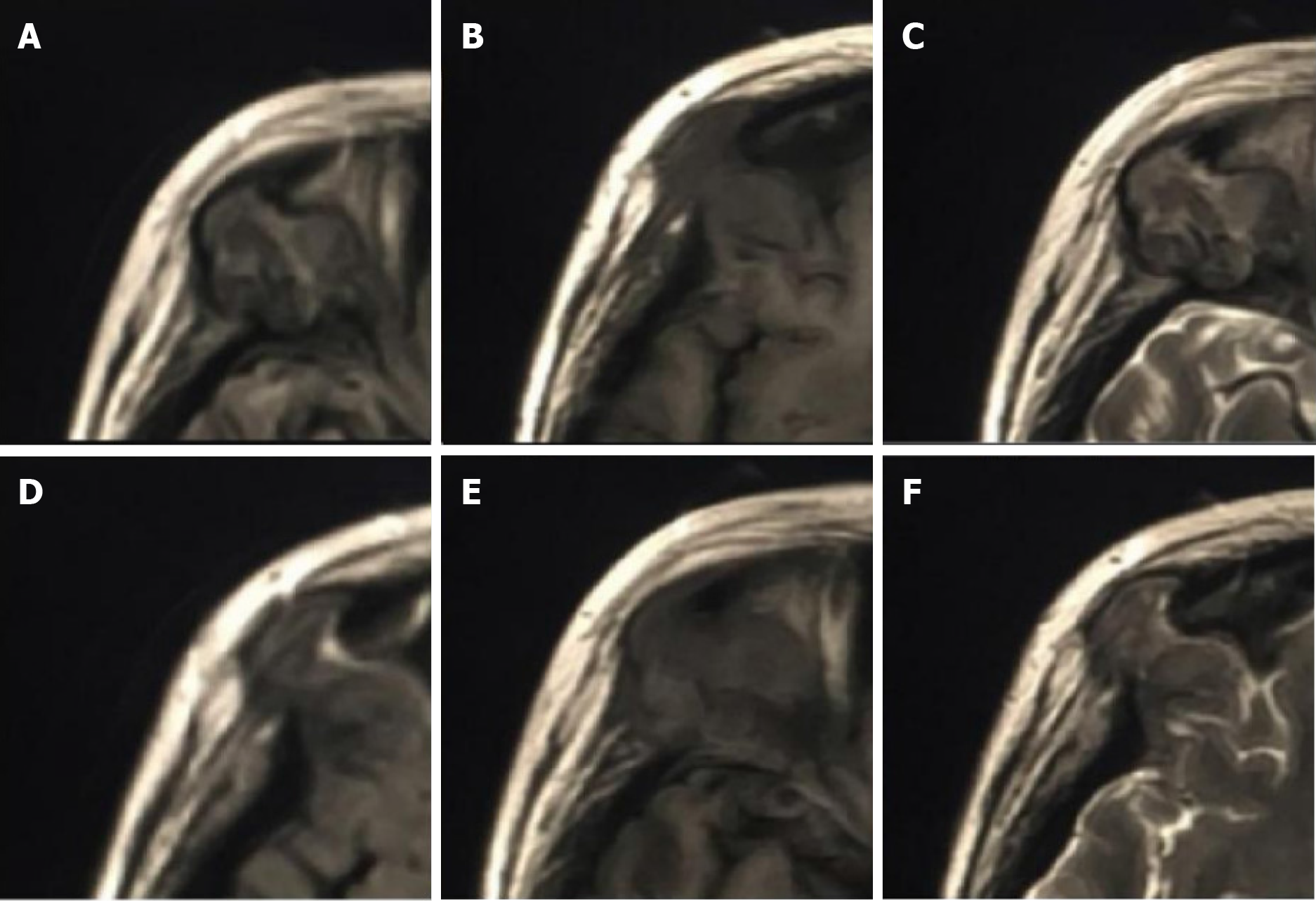
Figure 7 Orbital magnetic resonance imaging showed irregular bone destruction in the upper outer wall of the right eye orbit.
It was found with isotropic T1 and isotropic/slightly longer T2 signal shadows with isotropic signals in the fluid attenuated inversion recovery sequence. A-C: Representative the upper half of the lesion as seen on the axial fluid-attenuated inversion recovery, T2-weighted, and T1-weighted images; D-F: Representative the lower half of the lesion as seen on the axial fluid-attenuated inversion recovery, T2-weighted, and T1-weighted images.
- Citation: Zhang ZR, Chen F, Chen HJ. Multisystemic recurrent Langerhans cell histiocytosis misdiagnosed with chronic inflammation at the first diagnosis: A case report. World J Radiol 2024; 16(6): 232-240
- URL: https://www.wjgnet.com/1949-8470/full/v16/i6/232.htm
- DOI: https://dx.doi.org/10.4329/wjr.v16.i6.232


