Copyright
©The Author(s) 2024.
World J Radiol. Jun 28, 2024; 16(6): 232-240
Published online Jun 28, 2024. doi: 10.4329/wjr.v16.i6.232
Published online Jun 28, 2024. doi: 10.4329/wjr.v16.i6.232
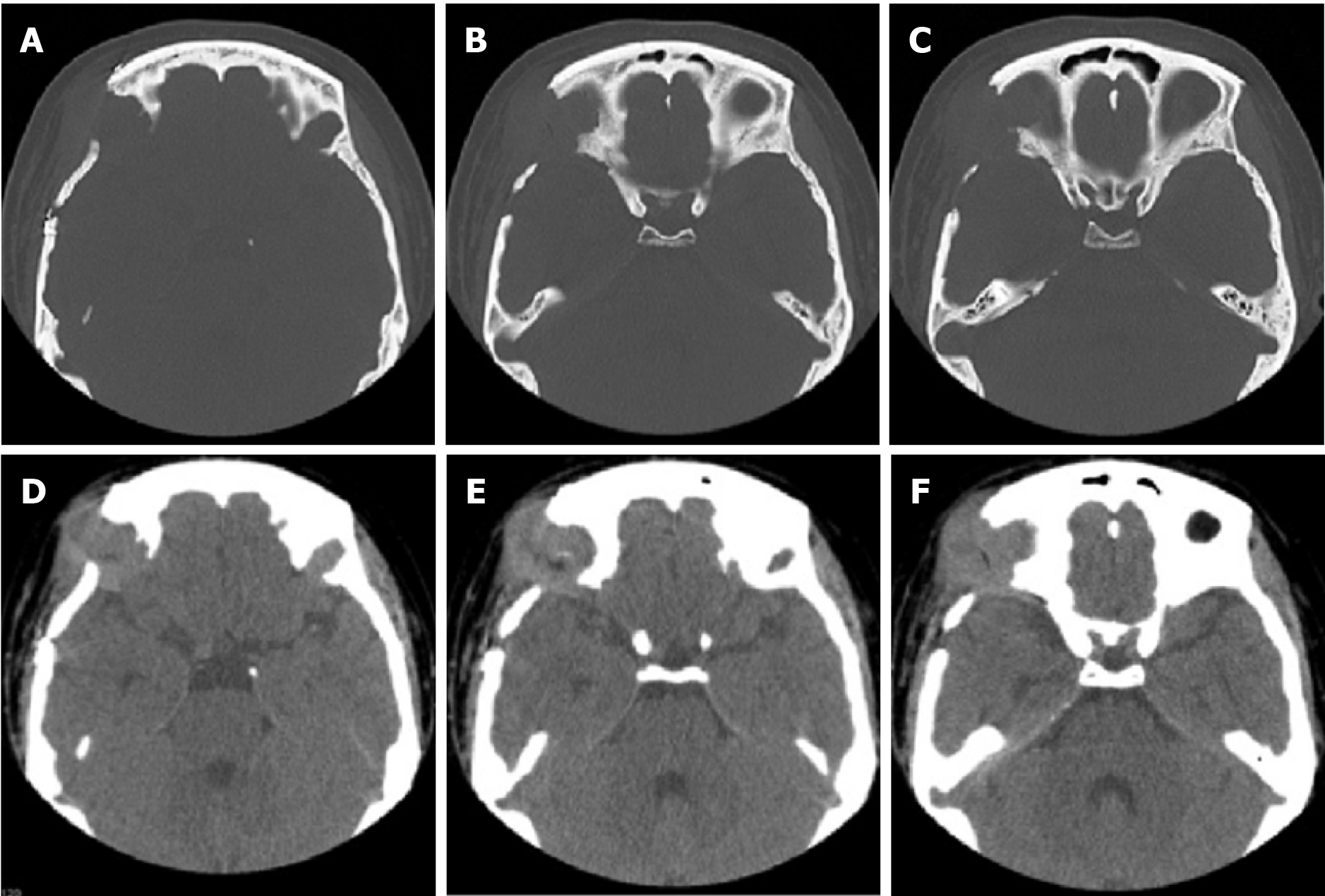
Figure 6 Orbital computed tomography showed irregular bone destruction in the upper outer wall of the right eye orbit combined with the formation of a surrounding soft tissue mass.
A-C: Representative axial bone views; D-F: Representative axial soft tissue views.
- Citation: Zhang ZR, Chen F, Chen HJ. Multisystemic recurrent Langerhans cell histiocytosis misdiagnosed with chronic inflammation at the first diagnosis: A case report. World J Radiol 2024; 16(6): 232-240
- URL: https://www.wjgnet.com/1949-8470/full/v16/i6/232.htm
- DOI: https://dx.doi.org/10.4329/wjr.v16.i6.232


