Copyright
©The Author(s) 2024.
World J Radiol. May 28, 2024; 16(5): 115-127
Published online May 28, 2024. doi: 10.4329/wjr.v16.i5.115
Published online May 28, 2024. doi: 10.4329/wjr.v16.i5.115
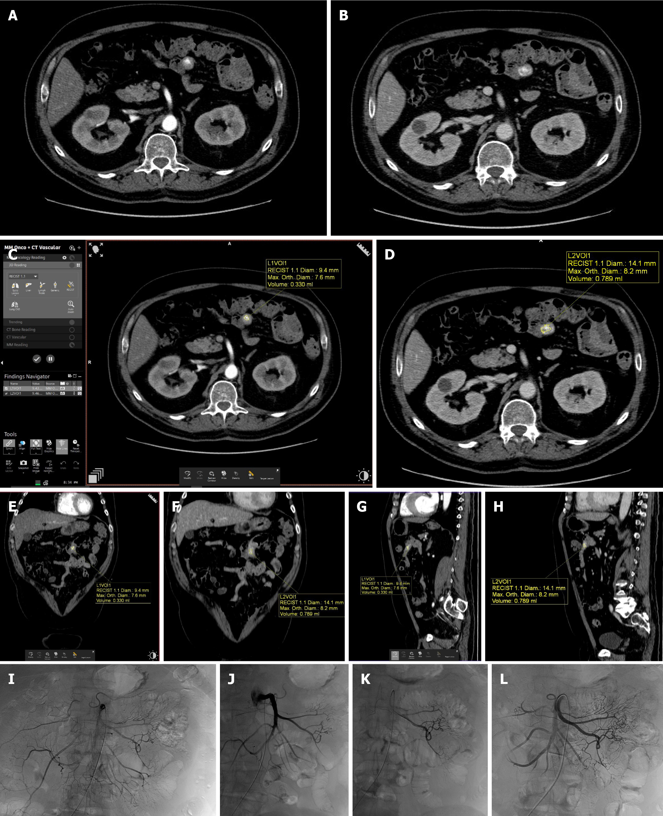
Figure 3 Example case of a 63-year-old man with jejunal gastrointestinal bleeding detected at computed tomography angiography and evaluated with a semiautomated software (bleeding rate: 5.
397 mL/min) and not confirmed by catheter angiography (non-concordance group). A: Computed tomography angiography (CTA) in the arterial phase showing jejunal gastrointestinal bleeding (GIB) on axial view; B: CTA in the venous phase showing jejunal GIB on axial view; C: GIB volume evaluation in the arterial phase of axial CTA with a semiautomatic software on a dedicated workstation. Bleeding volume was 0.330 mL; D: GIB volume evaluation in the venous phase of axial CTA with a semiautomatic software. Bleeding volume in the venous phase was 0.769 mL; E: GIB volume evaluation in the arterial phase of coronal CTA; F: GIB volume evaluation in the venous phase of coronal CTA; G: GIB volume evaluation in the arterial phase of sagittal CTA; H: GIB volume evaluation in the arterial phase of sagittal CTA; I: Catheter angiography of superior mesenteric artery showing no signs of bleeding; J-L: Superior mesenteric artery angiograms after multiple selective and super selective catheterisms of distal branches showing no signs of bleeding.
- Citation: Cacioppa LM, Floridi C, Bruno A, Rossini N, Valeri T, Borgheresi A, Inchingolo R, Cortese F, Novelli G, Felicioli A, Torresi M, Boscarato P, Ottaviani L, Giovagnoni A. Extravasated contrast volumetric assessment on computed tomography angiography in gastrointestinal bleeding: A useful predictor of positive angiographic findings. World J Radiol 2024; 16(5): 115-127
- URL: https://www.wjgnet.com/1949-8470/full/v16/i5/115.htm
- DOI: https://dx.doi.org/10.4329/wjr.v16.i5.115


