Copyright
©The Author(s) 2024.
World J Radiol. Apr 28, 2024; 16(4): 94-108
Published online Apr 28, 2024. doi: 10.4329/wjr.v16.i4.94
Published online Apr 28, 2024. doi: 10.4329/wjr.v16.i4.94
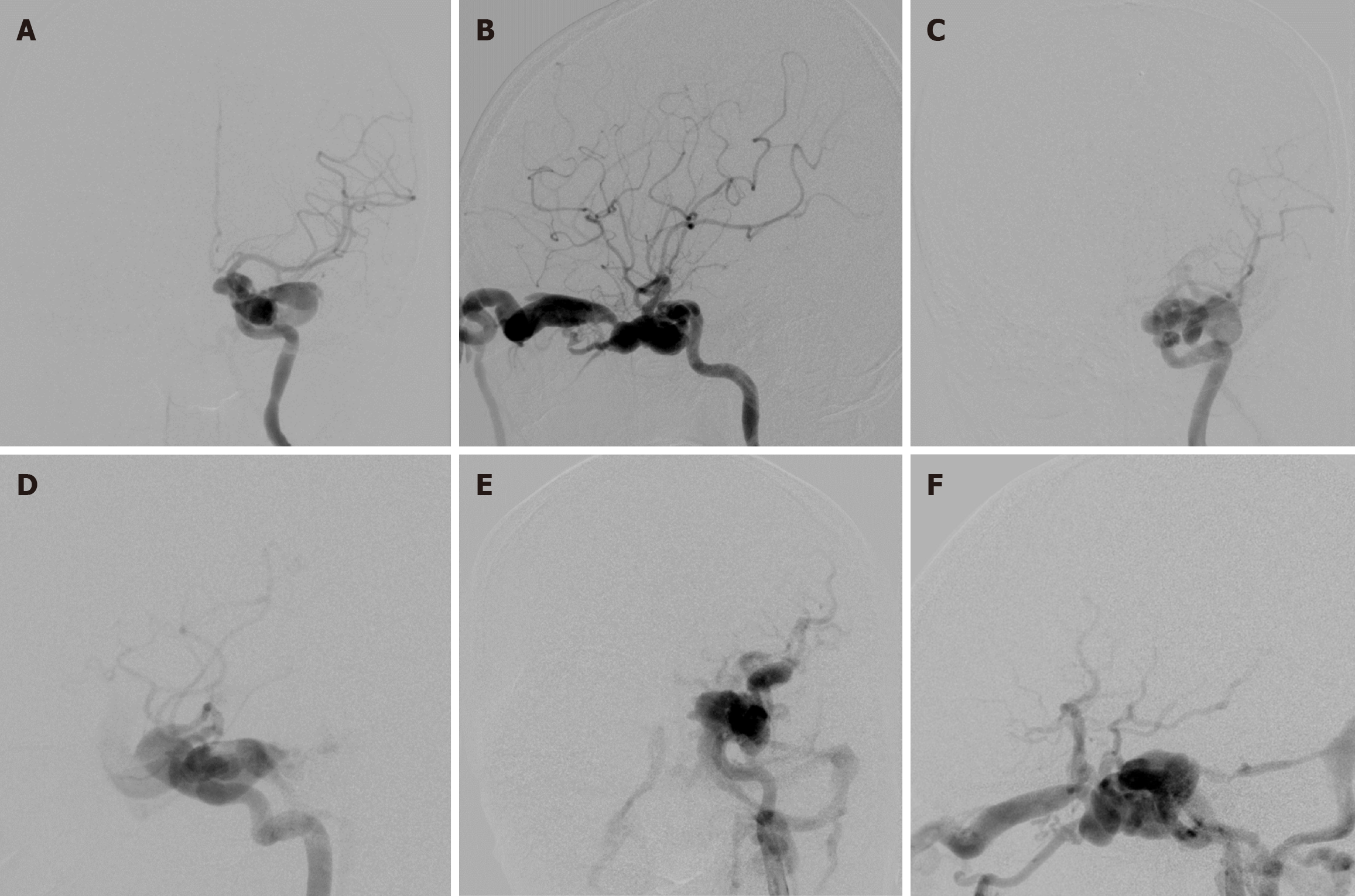
Figure 3 Classification of traumatic carotid-cavernous fistulas according to the fistula size.
A and B: Anteroposterior (AP) and lateral views of a small-sized fistula, which had good antegrade ipsilateral internal carotid artery (ICA) flow, represented by opacification of both the anterior cerebral artery (ACA) and middle cerebral artery (MCA); C and D: AP and lateral views of a medium-sized fistula, which had fair antegrade flow, represented by opacification of either the ACA or MCA; E and F: AP and lateral views of a large-sized fistula, which had poor antegrade flow, represented by opacification of neither the ACA nor MCA.
- Citation: Iampreechakul P, Wangtanaphat K, Chuntaroj S, Wattanasen Y, Hangsapruek S, Lertbutsayanukul P, Puthkhao P, Siriwimonmas S. Pseudoaneurysm formation following transarterial embolization of traumatic carotid-cavernous fistula with detachable balloon: An institutional cohort long-term study. World J Radiol 2024; 16(4): 94-108
- URL: https://www.wjgnet.com/1949-8470/full/v16/i4/94.htm
- DOI: https://dx.doi.org/10.4329/wjr.v16.i4.94


