Copyright
©The Author(s) 2024.
World J Radiol. Apr 28, 2024; 16(4): 82-93
Published online Apr 28, 2024. doi: 10.4329/wjr.v16.i4.82
Published online Apr 28, 2024. doi: 10.4329/wjr.v16.i4.82
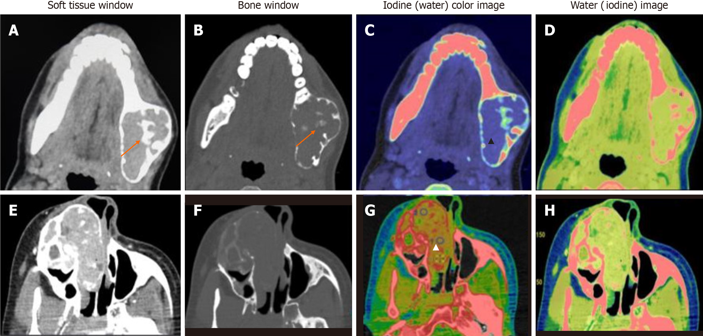
Figure 5 Mixed lytic-sclerotic lesions.
A-D: Central giant cell granuloma well-defined, mixed lytic-sclerotic buccolingual expansile lesion with a narrow zone of transition. Central ossific foci are seen (orange arrows). Iodine (water) material decomposition overlay images show a mild, homogeneous iodine concentration (blue region within the tumor - black arrowhead) (C). Water (Iodine) images show no cystic or necrotic areas (D); E-H: Ossifying fibroma well- defined expansile mass epicentered in the right maxilla, showing heterogeneous enhancement in its lytic soft tissue component with multiple sclerotic foci extending into the nasal cavity (E). The iodine image shows foci of increased iodine concentration (red areas - white arrowhead) (G). Lower iodine concentration and higher water concentration were seen in the latter, which suggested “other jaw tumor” as in this case.
- Citation: Viswanathan DJ, Bhalla AS, Manchanda S, Roychoudhury A, Mishra D, Mridha AR. Characterization of tumors of jaw: Additive value of contrast enhancement and dual-energy computed tomography. World J Radiol 2024; 16(4): 82-93
- URL: https://www.wjgnet.com/1949-8470/full/v16/i4/82.htm
- DOI: https://dx.doi.org/10.4329/wjr.v16.i4.82


