Copyright
©The Author(s) 2024.
World J Radiol. Apr 28, 2024; 16(4): 82-93
Published online Apr 28, 2024. doi: 10.4329/wjr.v16.i4.82
Published online Apr 28, 2024. doi: 10.4329/wjr.v16.i4.82
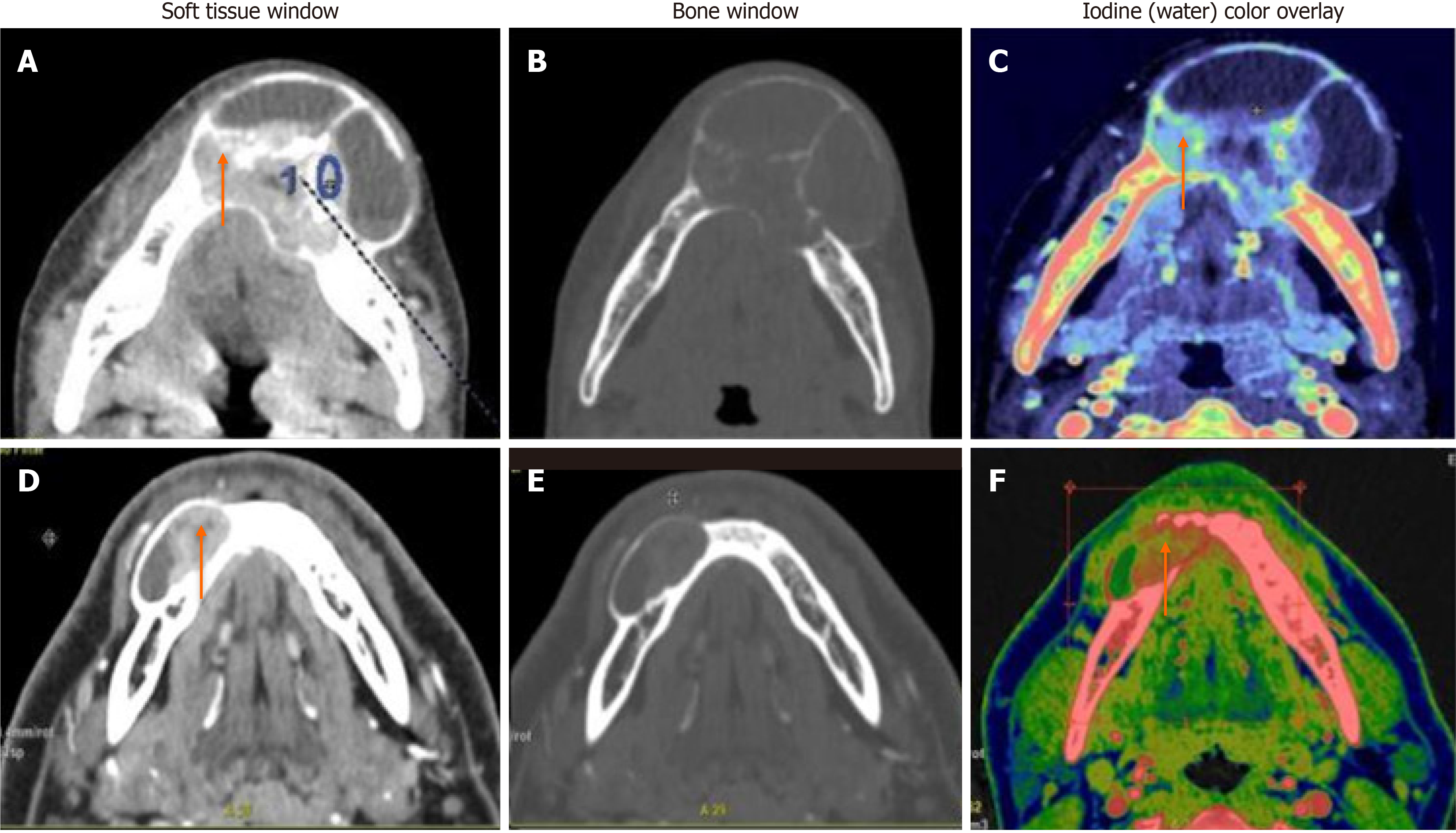
Figure 4 Multilocular solid-cystic lesions differentiated based on iodine concentration.
A-C: Central giant cell granuloma - A well-defined expansile multiloculated solid-cystic tumor in the left anterior mandible crossing the midline. Iodine images with a color overlay (C) showed an iodine concentration (IC) of 57 × 100 μg/cm3 of the solid enhancing part (orange arrow); D-F: Ameloblastoma - a well-defined lytic, unilocular, solid-cystic, expansile lesion with an enhancing soft tissue component (orange arrow). Iodine images with a color overlay (F) show increased iodine content (areas in red) within the soft tissue (IC of 23 × 100 μg/cm3).
- Citation: Viswanathan DJ, Bhalla AS, Manchanda S, Roychoudhury A, Mishra D, Mridha AR. Characterization of tumors of jaw: Additive value of contrast enhancement and dual-energy computed tomography. World J Radiol 2024; 16(4): 82-93
- URL: https://www.wjgnet.com/1949-8470/full/v16/i4/82.htm
- DOI: https://dx.doi.org/10.4329/wjr.v16.i4.82


