Copyright
©The Author(s) 2024.
World J Radiol. Apr 28, 2024; 16(4): 82-93
Published online Apr 28, 2024. doi: 10.4329/wjr.v16.i4.82
Published online Apr 28, 2024. doi: 10.4329/wjr.v16.i4.82
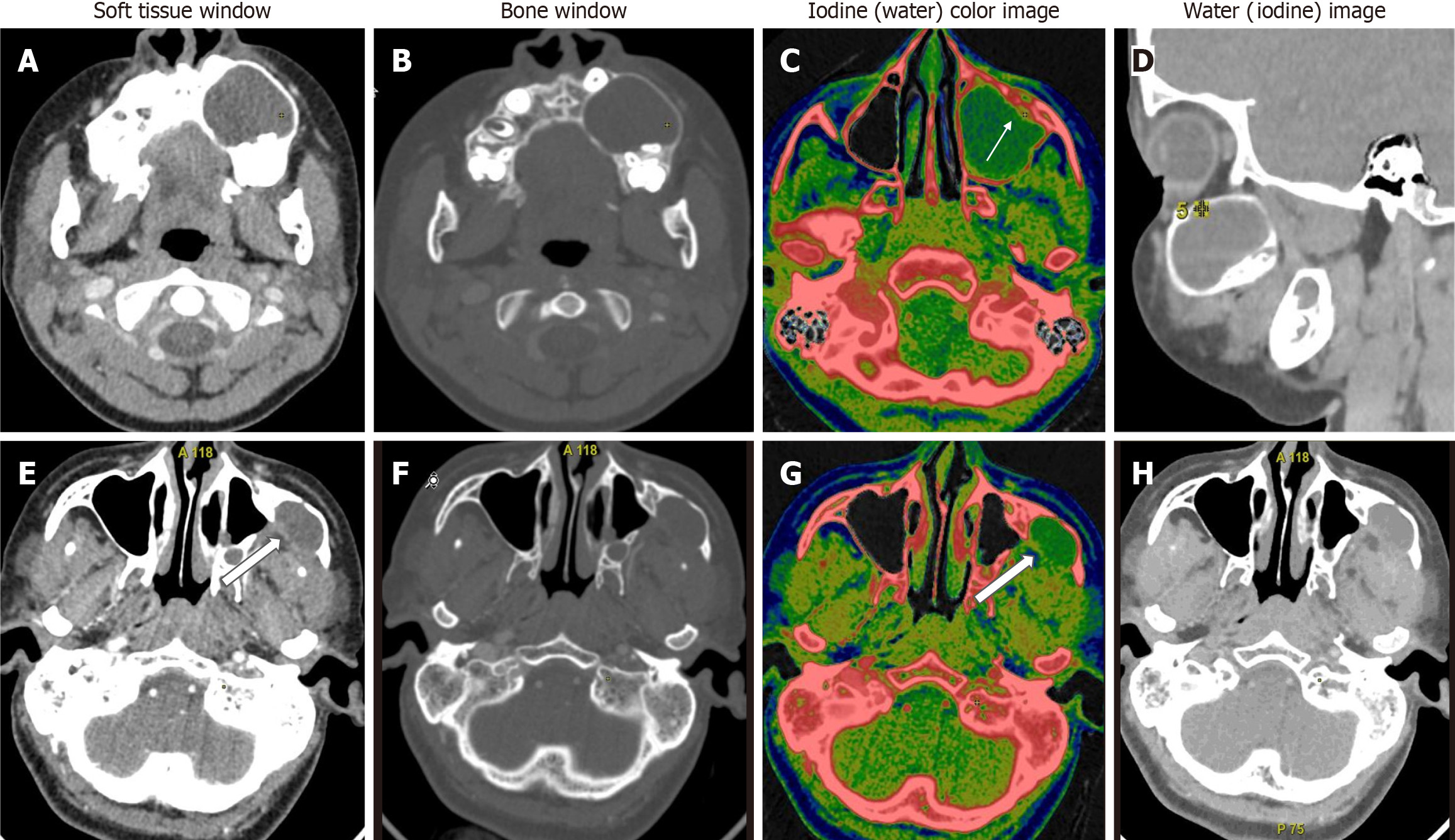
Figure 3 Unilocular lytic lesions differentiated based on water concentration.
A-D: Unilocular ameloblastoma - contrast-enhanced dual-energy computed tomography images show a well-defined lytic unilocular cystic, expansile lesion in the left maxilla with mild peripheral rim enhancement better appreciated on iodine colour overlay images (white arrow). Water (iodine) material decomposition (MD) images showed a WC of 986 μg/cm3 in the cystic component; E-H: Odontogenic keratocyst is also a well-defined lytic, unilocular cystic, expansile lesion in the left lateral wall of the maxillary sinus with a small enhancing mural component posteriorly (white open arrows). Water (Iodine) MD images revealed a water concentration of 1045 μg/cm3 in the cystic component. In this case, due to the paucity of soft tissue components, iodine concentration did not help; however, the water concentration of the cystic component differed significantly, aiding in the diagnosis.
- Citation: Viswanathan DJ, Bhalla AS, Manchanda S, Roychoudhury A, Mishra D, Mridha AR. Characterization of tumors of jaw: Additive value of contrast enhancement and dual-energy computed tomography. World J Radiol 2024; 16(4): 82-93
- URL: https://www.wjgnet.com/1949-8470/full/v16/i4/82.htm
- DOI: https://dx.doi.org/10.4329/wjr.v16.i4.82


