Copyright
©The Author(s) 2024.
World J Radiol. Apr 28, 2024; 16(4): 82-93
Published online Apr 28, 2024. doi: 10.4329/wjr.v16.i4.82
Published online Apr 28, 2024. doi: 10.4329/wjr.v16.i4.82
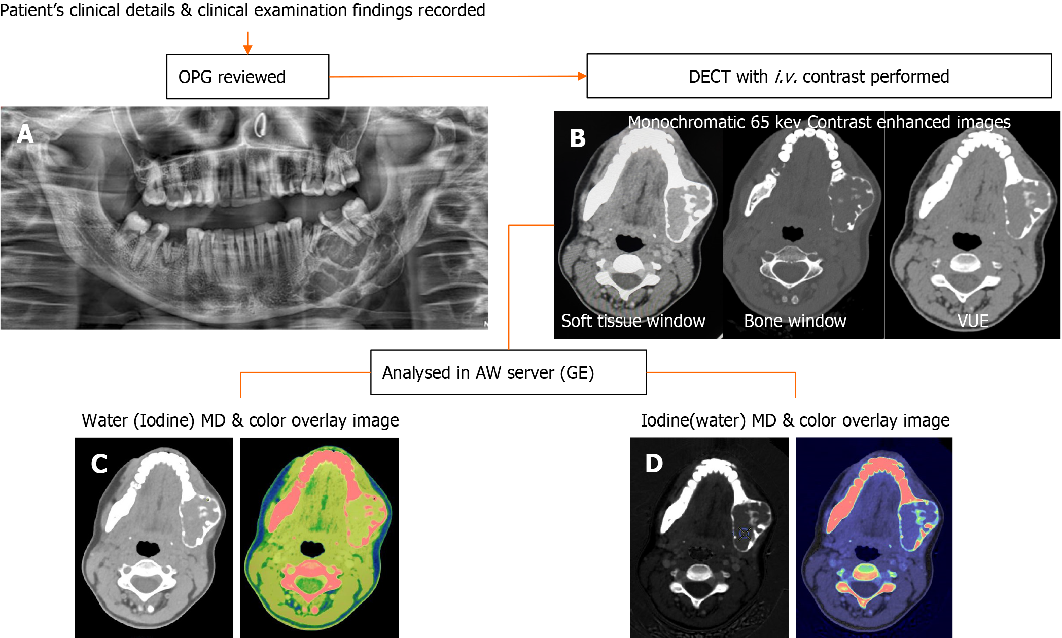
Figure 1 Workflow of patients undergoing dual-energy computed tomography Imaging.
A: Orthopantomogram images are reviewed first; B: Followed by dual-energy computed tomography (DECT) acquisition using intravenous non-ionic iodinated contrast; C: Water (Iodine) with color overlay; D: Iodine (water) with color overlay are the material density images reconstructed in the dedicated software for quantitative analysis. DECT: Dual-energy computed tomography; OPG: Orthopantomography; MD: Material decomposition.
- Citation: Viswanathan DJ, Bhalla AS, Manchanda S, Roychoudhury A, Mishra D, Mridha AR. Characterization of tumors of jaw: Additive value of contrast enhancement and dual-energy computed tomography. World J Radiol 2024; 16(4): 82-93
- URL: https://www.wjgnet.com/1949-8470/full/v16/i4/82.htm
- DOI: https://dx.doi.org/10.4329/wjr.v16.i4.82


