Copyright
©The Author(s) 2024.
World J Radiol. Mar 28, 2024; 16(3): 58-68
Published online Mar 28, 2024. doi: 10.4329/wjr.v16.i3.58
Published online Mar 28, 2024. doi: 10.4329/wjr.v16.i3.58
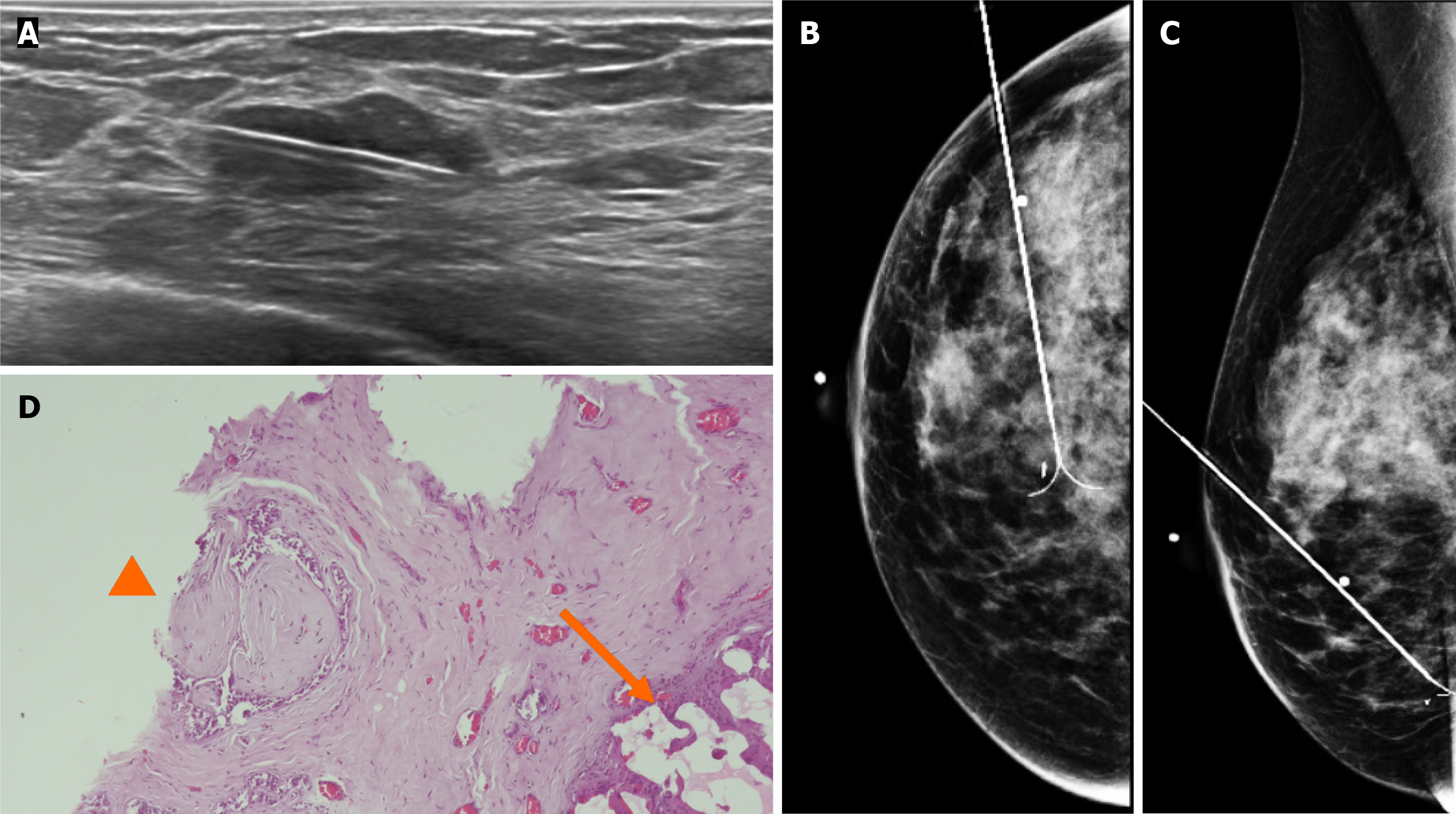
Figure 9 Gray-scale ultrasound images.
A: Preoperative guidewire is inserted percutaneously into the breast to localize the non-palpable conglomerate of nodules done under ultrasound; B and C: Craniocaudal and mediolateral oblique projection. The adequate position of the hook wire is confirmed in the target lesion; D: Histological slide of partial mastectomy. The site of the previous biopsy is seen (arrow). Also, fibroadenoma is shown which contains pleomorphic neoplastic cells and nucleolus with pleomorphic appearance (head arrow) that corresponds to ductal carcinoma in situ.
- Citation: Olivares-Antúnez Y, Dávila-Zablah YJ, Vázquez-Ávila JR, Gómez-Macías GS, Mireles-Aguilar MT, Garza-Montemayor ML. Ductal carcinoma in situ within a fibroadenoma: A case report and review of literature. World J Radiol 2024; 16(3): 58-68
- URL: https://www.wjgnet.com/1949-8470/full/v16/i3/58.htm
- DOI: https://dx.doi.org/10.4329/wjr.v16.i3.58


