Copyright
©The Author(s) 2024.
World J Radiol. Mar 28, 2024; 16(3): 58-68
Published online Mar 28, 2024. doi: 10.4329/wjr.v16.i3.58
Published online Mar 28, 2024. doi: 10.4329/wjr.v16.i3.58
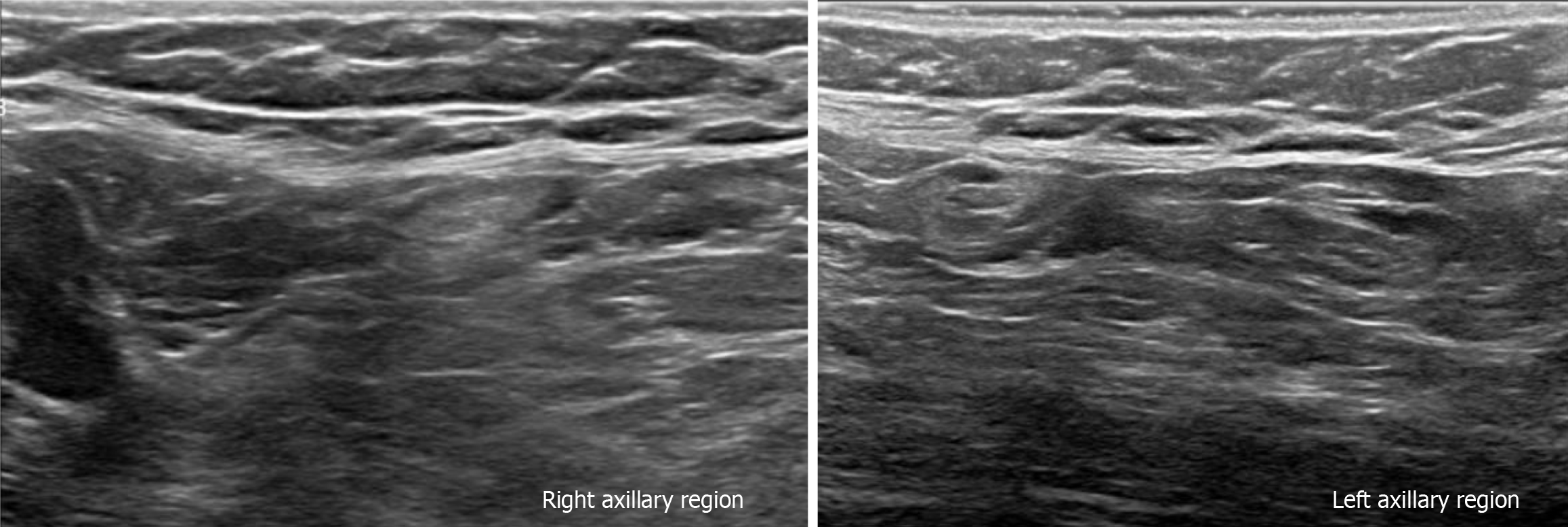
Figure 6 Gray-scale ultrasound images.
The bilateral axillary region shows lymph nodes with conservative morphology and fat hilum, with cortex of < 3 mm.
- Citation: Olivares-Antúnez Y, Dávila-Zablah YJ, Vázquez-Ávila JR, Gómez-Macías GS, Mireles-Aguilar MT, Garza-Montemayor ML. Ductal carcinoma in situ within a fibroadenoma: A case report and review of literature. World J Radiol 2024; 16(3): 58-68
- URL: https://www.wjgnet.com/1949-8470/full/v16/i3/58.htm
- DOI: https://dx.doi.org/10.4329/wjr.v16.i3.58


