Copyright
©The Author(s) 2024.
World J Radiol. Mar 28, 2024; 16(3): 40-48
Published online Mar 28, 2024. doi: 10.4329/wjr.v16.i3.40
Published online Mar 28, 2024. doi: 10.4329/wjr.v16.i3.40
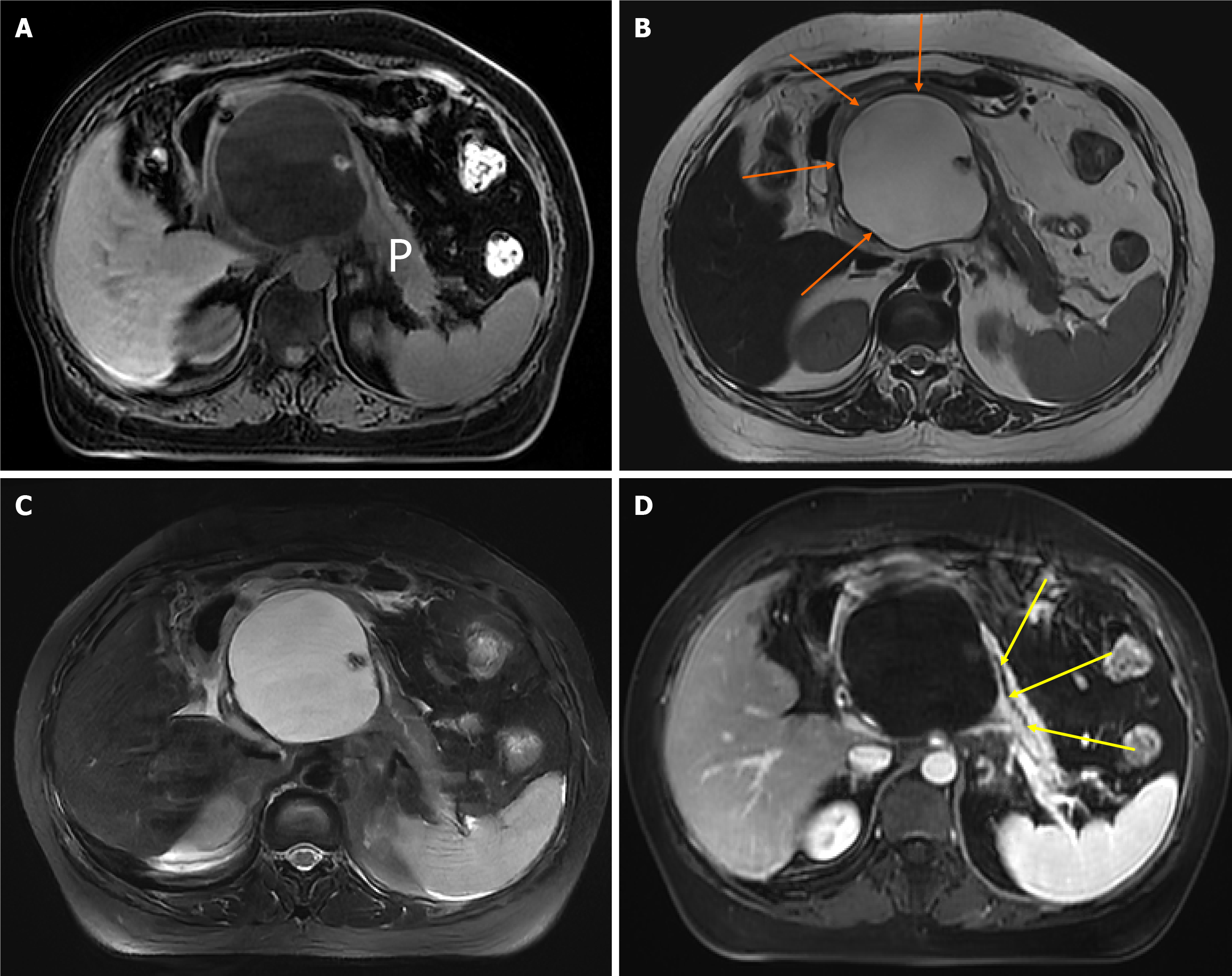
Figure 2 Chronic pancreatitis with large pancreatic cysts.
A 52-year-old woman with chronic pancreatitis presented with upper abdominal pain. The upper abdominal magnetic resonance images. A: Magnetic resonance imaging (MRI) fat-suppressed T1-weighted imaging shows a large pseudocyst of the head of the pancreas, which has a low signal; B: MRI T2-weighted imaging can show a clear boundary of pseudocyst (orange arrows) with a diameter of 7 cm × 11 cm, which has a high signal; C: MRI fat-suppressed T2-weighted imaging; D: MRI enhanced scanning venous phase shows the large pseudocyst has no enhancement as well as the displaced main pancreatic duct (yellow arrows). P: Pancreas.
- Citation: Feng Y, Song LJ, Xiao B. Chronic pancreatitis: Pain and computed tomography/magnetic resonance imaging findings. World J Radiol 2024; 16(3): 40-48
- URL: https://www.wjgnet.com/1949-8470/full/v16/i3/40.htm
- DOI: https://dx.doi.org/10.4329/wjr.v16.i3.40


