Copyright
©The Author(s) 2024.
World J Radiol. Mar 28, 2024; 16(3): 40-48
Published online Mar 28, 2024. doi: 10.4329/wjr.v16.i3.40
Published online Mar 28, 2024. doi: 10.4329/wjr.v16.i3.40
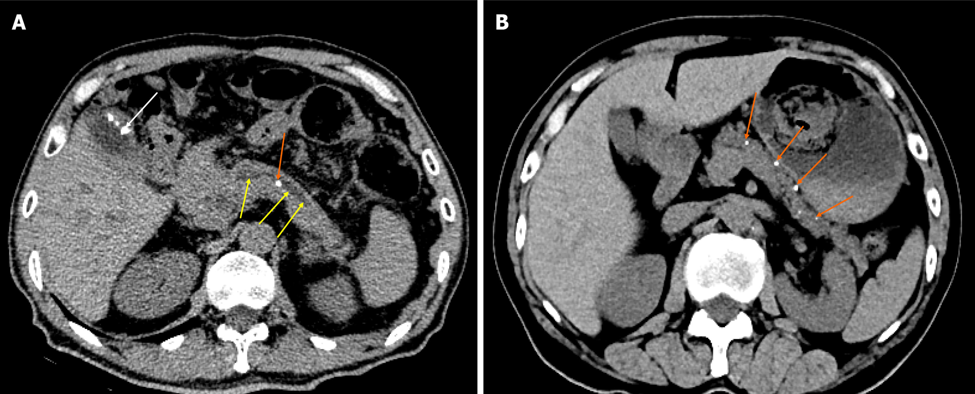
Figure 1 Chronic pancreatitis.
A: Chronic pancreatitis with main pancreatic duct stone and main pancreatic duct dilatation. A 58-year-old man with chronic pancreatitis presented with no pain. The abdominal computed tomography (CT) scan represents the main pancreatic duct stone (orange arrow), the dilated main pancreatic duct (yellow arrows) with a diameter of 0.5 cm, as well as the combined gallbladder multiple stones (white arrow); B: Chronic pancreatitis with pancreatic parenchymal atrophy and multiple pancreatic calcifications. A 69-year-old man with chronic pancreatitis. The abdominal CT scan shows a decrease in pancreatic volume, parenchymal atrophy, and multiple calcifications in the pancreatic parenchyma (orange arrows).
- Citation: Feng Y, Song LJ, Xiao B. Chronic pancreatitis: Pain and computed tomography/magnetic resonance imaging findings. World J Radiol 2024; 16(3): 40-48
- URL: https://www.wjgnet.com/1949-8470/full/v16/i3/40.htm
- DOI: https://dx.doi.org/10.4329/wjr.v16.i3.40


