Copyright
©The Author(s) 2024.
World J Radiol. Dec 28, 2024; 16(12): 771-781
Published online Dec 28, 2024. doi: 10.4329/wjr.v16.i12.771
Published online Dec 28, 2024. doi: 10.4329/wjr.v16.i12.771
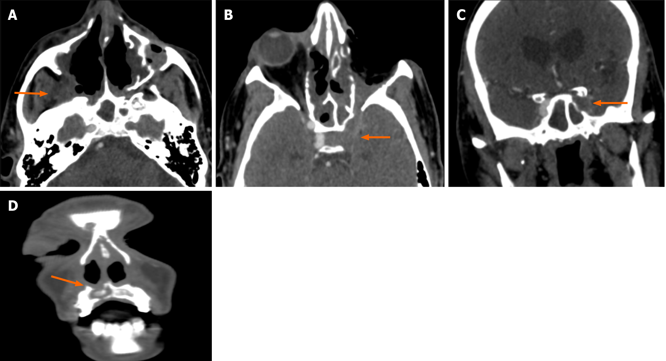
Figure 4 Repeat imaging after 10 weeks of patient in Figure 3 (known case of rhinoorbitocerebral mucormycosis with severe disease and post left exenteration).
A and B: Axial contrast-enhanced computed tomography images (A and B) reveal soft tissue infiltrating the right posterior antral space (arrow in A) and soft tissue in the left orbit with contiguous extension to the left orbital apex and left cavernous sinus (arrow in B). Left post-exenteration status can be seen; C and D: Coronal images show soft tissue in the left cavernous sinus with the non-opacified cavernous segment of the left internal carotid artery suggesting its occlusion (arrow in C). There is also evidence of maxillary alveolus destruction and sequestrum formation (arrow in D). This repeat imaging shows radiological worsening post-treatment (non-responder).
- Citation: Manchanda S, Bhalla AS, Nair AD, Sikka K, Verma H, Thakar A, Kakkar A, Khan MA. Proposed computed tomography severity index for the evaluation of invasive fungal sinusitis: Preliminary results. World J Radiol 2024; 16(12): 771-781
- URL: https://www.wjgnet.com/1949-8470/full/v16/i12/771.htm
- DOI: https://dx.doi.org/10.4329/wjr.v16.i12.771


