Copyright
©The Author(s) 2024.
World J Radiol. Dec 28, 2024; 16(12): 771-781
Published online Dec 28, 2024. doi: 10.4329/wjr.v16.i12.771
Published online Dec 28, 2024. doi: 10.4329/wjr.v16.i12.771
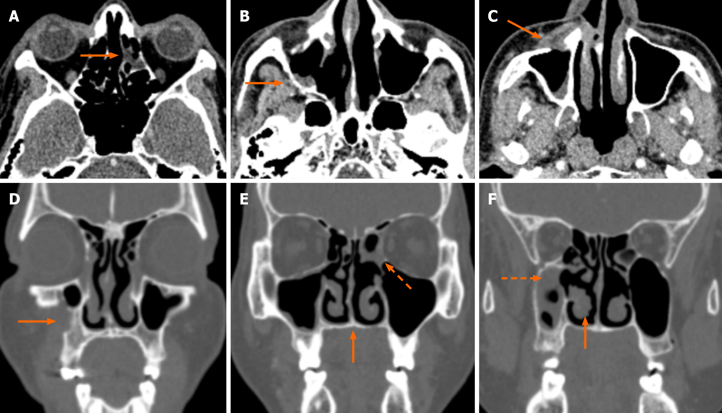
Figure 2 Mild disease with computed tomography severity index 2 (67-year-old male, coronavirus disease 2019 positive with mild headache and right-sided facial pain).
The patient was a known diabetic with an HbA1C of 8.5. Microbiological evaluation revealed both Aspergillus sp. and Rhizopus sp. A-C: Axial computed tomography (CT) images show minimal soft tissue within the left ethmoid sinus (A) and right maxillary sinus infiltrating the adjacent right posterior retroantral fat (B). The bilateral eye globes and orbits are normal. There is e/o erosion in the anterior maxillary wall with soft tissue infiltrating the preantral space (C); D-F: Coronal bone window CT images. Figure D shows bone erosion in the anterior maxilla (arrow). There is evidence of bilateral disease with mild mucosal disease in the right maxillary sinus (dotted arrow in F) and left ethmoid sinus (dotted arrow in E) with mucosal thickening of the right inferior turbinate (arrow in F). The hard palate is intact (arrow in E). The total summated CT severity index for this case is 2 - mild disease (1 point for involvement of right maxillary sinus and ethmoid sinus, 1 point for the involvement of anterior and posterior periantral fat). The patient was given intravenous amphotericin to which the patient responded well. Repeat imaging was not done in this case.
- Citation: Manchanda S, Bhalla AS, Nair AD, Sikka K, Verma H, Thakar A, Kakkar A, Khan MA. Proposed computed tomography severity index for the evaluation of invasive fungal sinusitis: Preliminary results. World J Radiol 2024; 16(12): 771-781
- URL: https://www.wjgnet.com/1949-8470/full/v16/i12/771.htm
- DOI: https://dx.doi.org/10.4329/wjr.v16.i12.771


