Copyright
©The Author(s) 2024.
World J Radiol. Dec 28, 2024; 16(12): 722-748
Published online Dec 28, 2024. doi: 10.4329/wjr.v16.i12.722
Published online Dec 28, 2024. doi: 10.4329/wjr.v16.i12.722
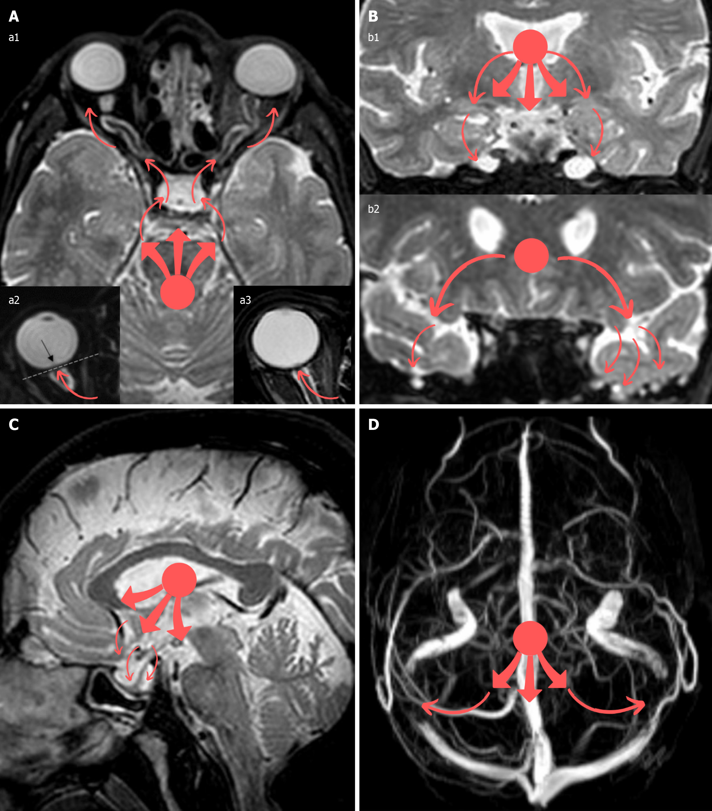
Figure 15 Idiopathic intracranial hypertension illustration.
A: In this illustration, the red dot in all panels represents an assumption of the supposed reference point from where the increased intracranial forces arise, while all the red arrows represent the outward direction of this force, which is attempting to release itself. As a result of intracranial hypertension, the force (red dot) is directed (red arrows) towards the optic nerves and their sheaths, causing their distension and tortuosity (panel a1), flattening of the posterior aspect of the globes (panel a2), and protrusion of the optic nerve head within the globe (panel a3); B: In a similar manner, the Meckel’s caves may be distended in an attempt to accommodate the excess cerebrospinal fluid pressure (panel b1), and meningoceles may also be created, mainly in the sphenoid bone wings and temporal bones, through bony clefts (panel b2); C: The pituitary gland may be stretched downwards against the expanded sella turcica (panel C), thus creating the empty or partially empty sella appearance; D: Compression of the transverse venous sinuses against the cranial bony cavity can result in stenosis of these veins (panel D) (although stenosis may preexist, causing an increase in intracranial pressure secondarily). This illustration/mechanism is only hypothetical and solely described in a rudimentary way for educational purposes to assist in memorizing the neuroimaging finding’s end results; besides, as previously mentioned, the exact pathogenesis of idiopathic intracranial hypertension (IIH) is complex and multifactorial. Neuroimaging findings may be promising clues for IIH diagnosis, although their absence does not rule it out. Various combinations of the related neuroimaging findings described may be encountered in IIH cases, and the radiologists should be aware of them to assist in the proper and timely diagnosis of the condition. Nonetheless, the role of the radiologists will primarily entail the exclusion of other intracranial pathologies hindering alternative diagnosis.
- Citation: Arkoudis NA, Davoutis E, Siderakis M, Papagiannopoulou G, Gouliopoulos N, Tsetsou I, Efthymiou E, Moschovaki-Zeiger O, Filippiadis D, Velonakis G. Idiopathic intracranial hypertension: Imaging and clinical fundamentals. World J Radiol 2024; 16(12): 722-748
- URL: https://www.wjgnet.com/1949-8470/full/v16/i12/722.htm
- DOI: https://dx.doi.org/10.4329/wjr.v16.i12.722


