Copyright
©The Author(s) 2024.
World J Radiol. Dec 28, 2024; 16(12): 722-748
Published online Dec 28, 2024. doi: 10.4329/wjr.v16.i12.722
Published online Dec 28, 2024. doi: 10.4329/wjr.v16.i12.722
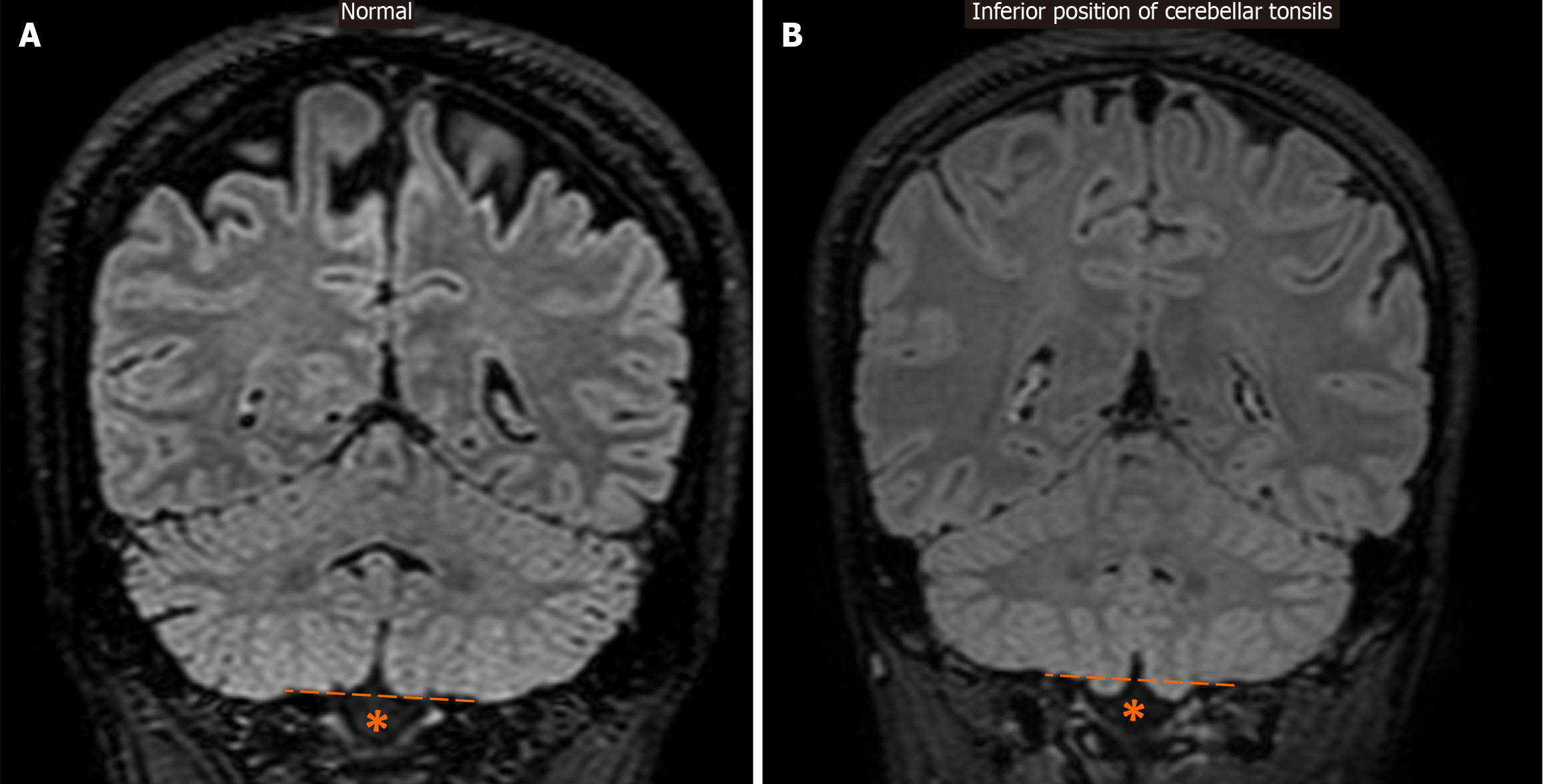
Figure 14 Inferior position of cerebellar tonsils.
A: For reference, a coronal fluid-attenuated inversion recovery image is provided, which displays normally positioned cerebellar tonsils above the level of the foramen magnum (*), as indicated by the dashed line; B: Coronal fluid-attenuated inversion recovery image demonstrates only mild ectopic localization of the inferior part of the cerebellar tonsils, just below their expected level (dashed line) and through the foramen magnum (*).
- Citation: Arkoudis NA, Davoutis E, Siderakis M, Papagiannopoulou G, Gouliopoulos N, Tsetsou I, Efthymiou E, Moschovaki-Zeiger O, Filippiadis D, Velonakis G. Idiopathic intracranial hypertension: Imaging and clinical fundamentals. World J Radiol 2024; 16(12): 722-748
- URL: https://www.wjgnet.com/1949-8470/full/v16/i12/722.htm
- DOI: https://dx.doi.org/10.4329/wjr.v16.i12.722


