Copyright
©The Author(s) 2024.
World J Radiol. Dec 28, 2024; 16(12): 722-748
Published online Dec 28, 2024. doi: 10.4329/wjr.v16.i12.722
Published online Dec 28, 2024. doi: 10.4329/wjr.v16.i12.722
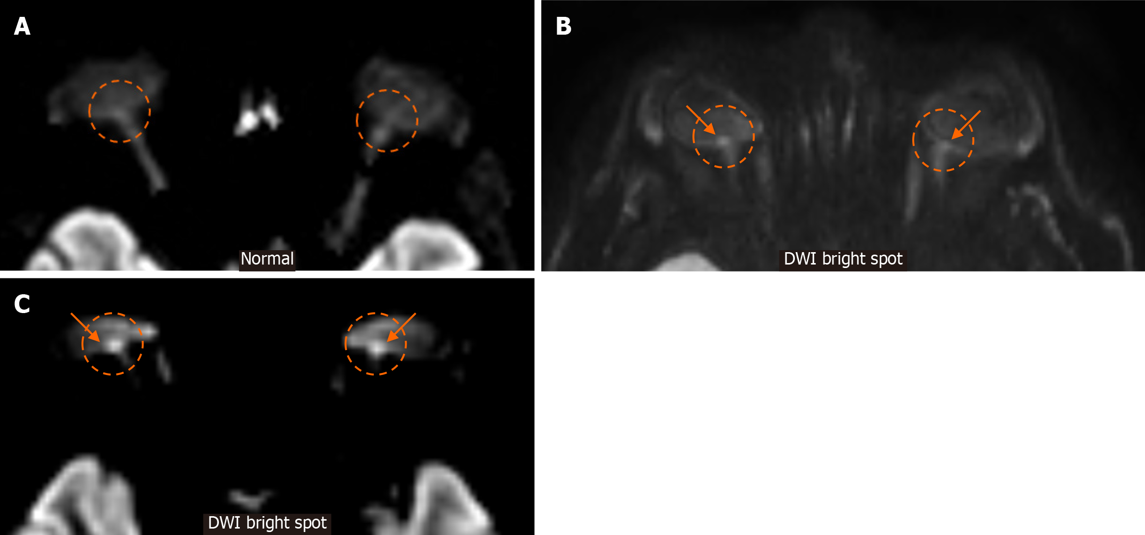
Figure 11 Diffusion-weighted imaging bright spot at fundus.
A: For reference, we provide an axial image of a diffusion-weighted imaging (DWI) magnetic resonance sequence from a normal patient. Note the absence of signal intensity in the posterior aspect of both globes in the expected location of the optic nerve heads (dashed circles), which corresponds to the expected normal appearance; B and C: Display axial DWI images in different patients with idiopathic intracranial hypertension. Note the obvious abnormal DWI signal hyperintensity in the optic nerve heads (arrows in dashed circles). DWI: Diffusion-weighted imaging.
- Citation: Arkoudis NA, Davoutis E, Siderakis M, Papagiannopoulou G, Gouliopoulos N, Tsetsou I, Efthymiou E, Moschovaki-Zeiger O, Filippiadis D, Velonakis G. Idiopathic intracranial hypertension: Imaging and clinical fundamentals. World J Radiol 2024; 16(12): 722-748
- URL: https://www.wjgnet.com/1949-8470/full/v16/i12/722.htm
- DOI: https://dx.doi.org/10.4329/wjr.v16.i12.722


