Copyright
©The Author(s) 2024.
World J Radiol. Dec 28, 2024; 16(12): 722-748
Published online Dec 28, 2024. doi: 10.4329/wjr.v16.i12.722
Published online Dec 28, 2024. doi: 10.4329/wjr.v16.i12.722
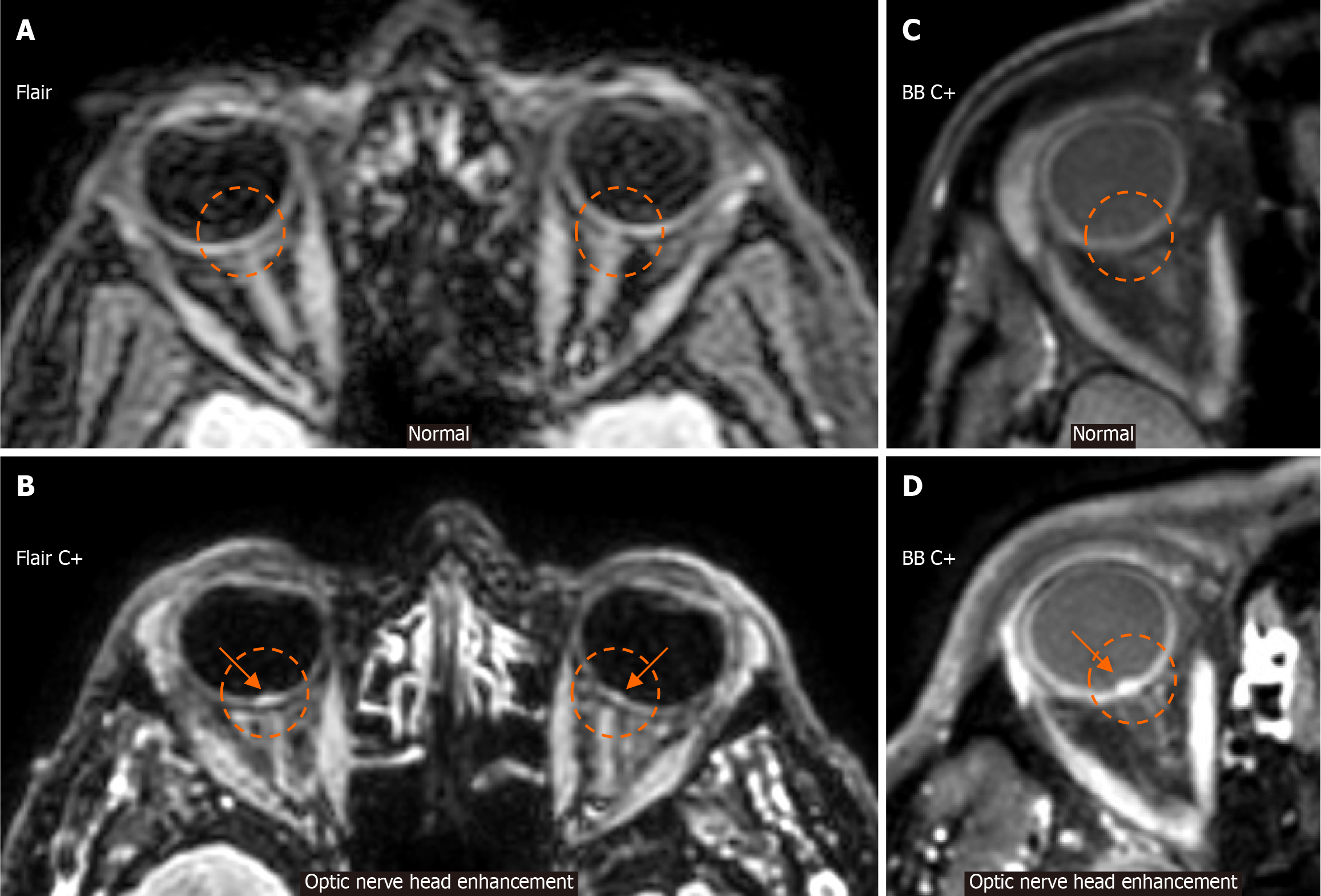
Figure 9 Optic nerve head enhancement.
A: An axial fluid-attenuated inversion recovery image at the level of the orbits from a patient without idiopathic intracranial hypertension (IIH) signs or symptoms is provided for reference. Note the absence of signal intensity in the posterior aspect of both globes in the expected location of the optic nerve heads (ONHs) (dashed circles), corresponding to the expected normal appearance; B: An axial fluid-attenuated inversion recovery image, following intravenous gadolinium injection (C+), at the level of the orbits in a patient with IIH is provided. Note the signal hyperintensity in the posterior aspect of both globes in the expected location of the ONHs, compatible with ONH enhancement (arrows within dashed circles), which is more evident on the right than on the left ONH; C: An axial black blood image, following intravenous gadolinium injection (C+), at the level of the orbits from a normal patient is provided for reference. Observe the absence of signal intensity in the posterior aspect of the globe in the expected location of the ONH (dashed circle); D: An axial black blood image, following intravenous gadolinium injection (C+), at the level of the orbits from a patient with IIH is provided. Note the signal hyperintensity in the posterior aspect of the globe in the expected location of the ONH, compatible with ONH enhancement (arrow within the dashed circle). BB: Black blood; Flair: Fluid-attenuated inversion recovery.
- Citation: Arkoudis NA, Davoutis E, Siderakis M, Papagiannopoulou G, Gouliopoulos N, Tsetsou I, Efthymiou E, Moschovaki-Zeiger O, Filippiadis D, Velonakis G. Idiopathic intracranial hypertension: Imaging and clinical fundamentals. World J Radiol 2024; 16(12): 722-748
- URL: https://www.wjgnet.com/1949-8470/full/v16/i12/722.htm
- DOI: https://dx.doi.org/10.4329/wjr.v16.i12.722


