Copyright
©The Author(s) 2024.
World J Radiol. Dec 28, 2024; 16(12): 722-748
Published online Dec 28, 2024. doi: 10.4329/wjr.v16.i12.722
Published online Dec 28, 2024. doi: 10.4329/wjr.v16.i12.722
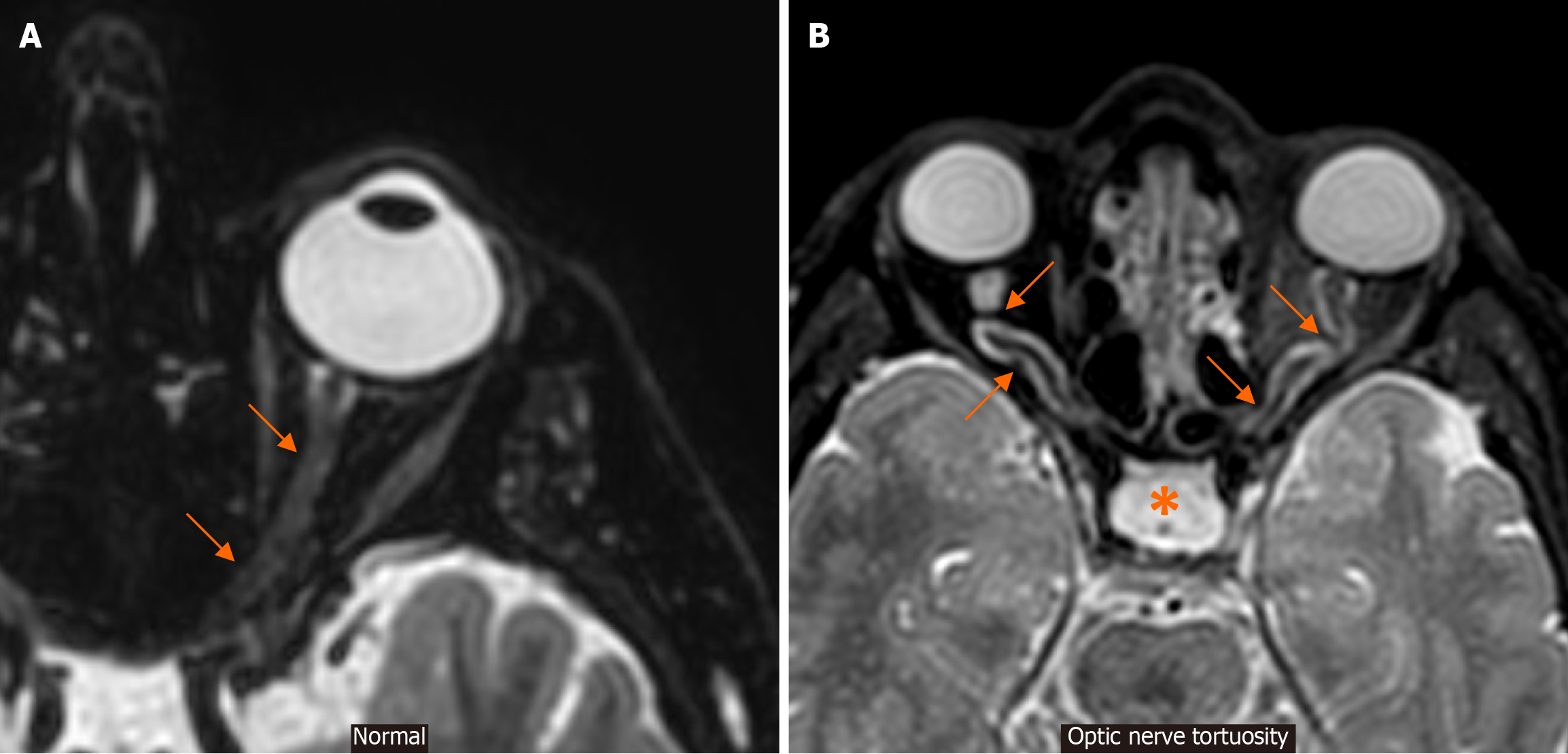
Figure 7 Optic nerve tortuosity.
A: An axial T2-weighted magnetic resonance image at the level of the globe and optic nerve in a patient with no intracranial hypertension signs or symptoms is provided for reference. Note the course of this normal-appearing optic nerve (arrows); B: An axial T2-weighted magnetic resonance image at the level of the globe and optic nerve in a patient with signs and symptoms of idiopathic intracranial hypertension demonstrates obvious tortuosity of the optic nerves in their intraorbital segments (arrows). Also note an empty sella appearance (*) and optic nerve sheath distension, representing additional signs supporting this diagnosis.
- Citation: Arkoudis NA, Davoutis E, Siderakis M, Papagiannopoulou G, Gouliopoulos N, Tsetsou I, Efthymiou E, Moschovaki-Zeiger O, Filippiadis D, Velonakis G. Idiopathic intracranial hypertension: Imaging and clinical fundamentals. World J Radiol 2024; 16(12): 722-748
- URL: https://www.wjgnet.com/1949-8470/full/v16/i12/722.htm
- DOI: https://dx.doi.org/10.4329/wjr.v16.i12.722


