Copyright
©The Author(s) 2024.
World J Radiol. Nov 28, 2024; 16(11): 668-677
Published online Nov 28, 2024. doi: 10.4329/wjr.v16.i11.668
Published online Nov 28, 2024. doi: 10.4329/wjr.v16.i11.668
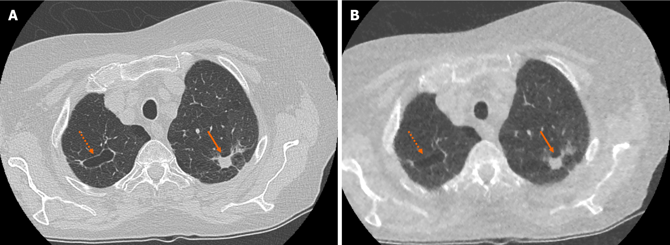
Figure 2 Example of accurate pulmonary nodule characterisation on ultra-low-dose computed tomography chest.
A: Selected axial slice of a standard dose computed tomography (CT) chest presented in lung windows with a spiculated solid pulmonary nodule with pleural tethering in the apico-posterior segment of the left upper lobe (solid arrow) and a parenchymal cyst in the apical segment of the right upper lobe (dashed arrow); B: Selected axial slice in the same patient presented in lung windows at the same level highlights the ability of ultra-low-dose CT chest with model-based iterative reconstruction to correctly characterise pulmonary nodule features such as spiculation, tethering and cavitation.
- Citation: O'Regan PW, Harold-Barry A, O'Mahony AT, Crowley C, Joyce S, Moore N, O'Connor OJ, Henry MT, Ryan DJ, Maher MM. Ultra-low-dose chest computed tomography with model-based iterative reconstruction in the analysis of solid pulmonary nodules: A prospective study. World J Radiol 2024; 16(11): 668-677
- URL: https://www.wjgnet.com/1949-8470/full/v16/i11/668.htm
- DOI: https://dx.doi.org/10.4329/wjr.v16.i11.668


