Copyright
©The Author(s) 2024.
World J Radiol. Nov 28, 2024; 16(11): 657-667
Published online Nov 28, 2024. doi: 10.4329/wjr.v16.i11.657
Published online Nov 28, 2024. doi: 10.4329/wjr.v16.i11.657
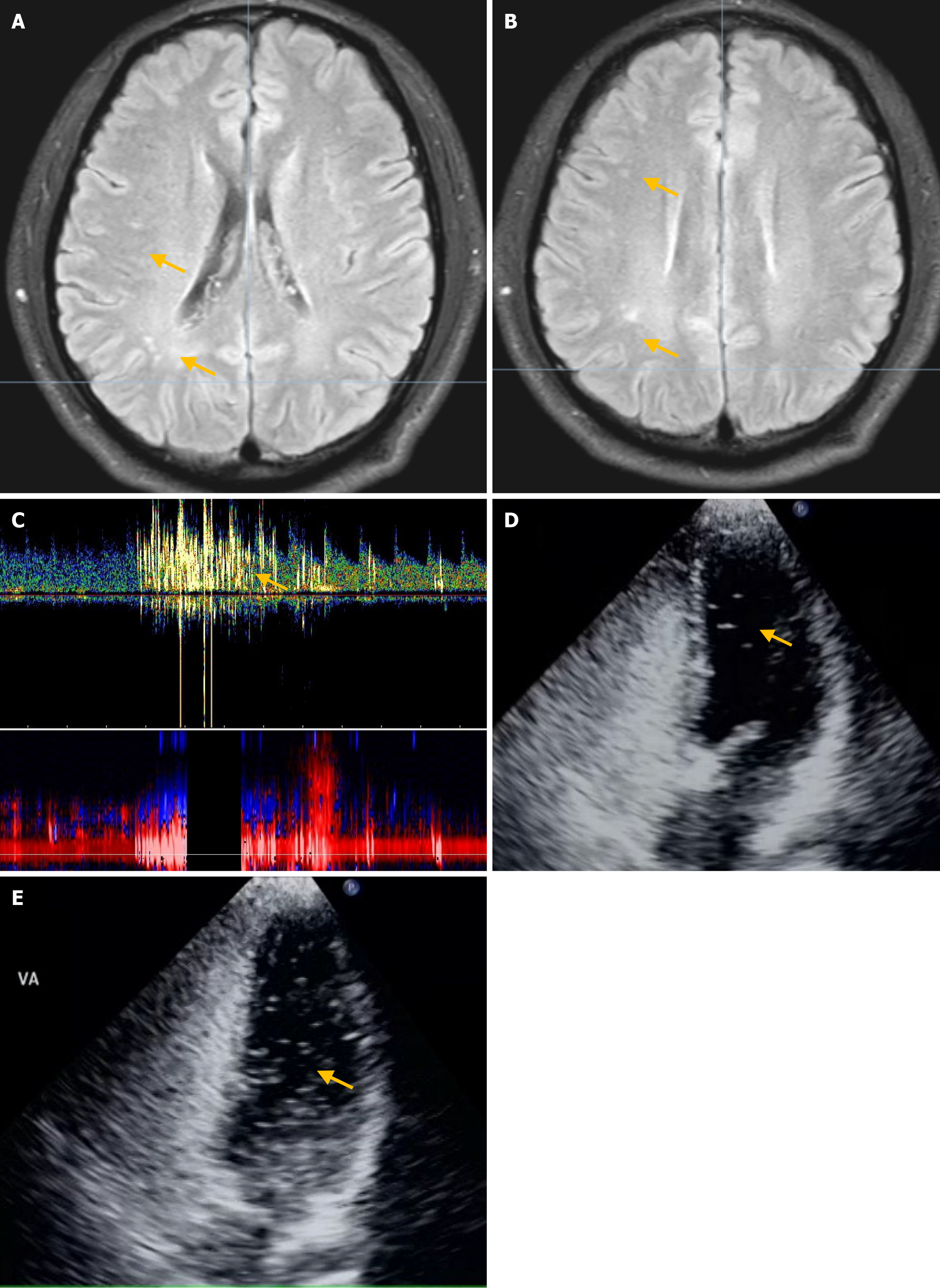
Figure 4 Magnetic resonance imaging of the brain.
A and B: Brain magnetic resonance imaging identified a few small ischemic foci in bilateral subcortical areas (arrows); C: At rest, contrast transcranial Doppler shows a small right-to-left shunt (RLS), whereas after the Valsalva maneuver (VM) it shows a large RLS; D and E: Contrast transthoracic echocardiography shows a small and a moderate RLS at rest and after the VM, respectively.
- Citation: Yao MJ, Zhao YY, Deng SP, Xiong HH, Wang J, Ren LJ, Cao LM. Right-to-left shunt detection via synchronized contrast transcranial Doppler combined with contrast transthoracic echocardiography: A preliminary study. World J Radiol 2024; 16(11): 657-667
- URL: https://www.wjgnet.com/1949-8470/full/v16/i11/657.htm
- DOI: https://dx.doi.org/10.4329/wjr.v16.i11.657


