Copyright
©The Author(s) 2024.
World J Radiol. Nov 28, 2024; 16(11): 657-667
Published online Nov 28, 2024. doi: 10.4329/wjr.v16.i11.657
Published online Nov 28, 2024. doi: 10.4329/wjr.v16.i11.657
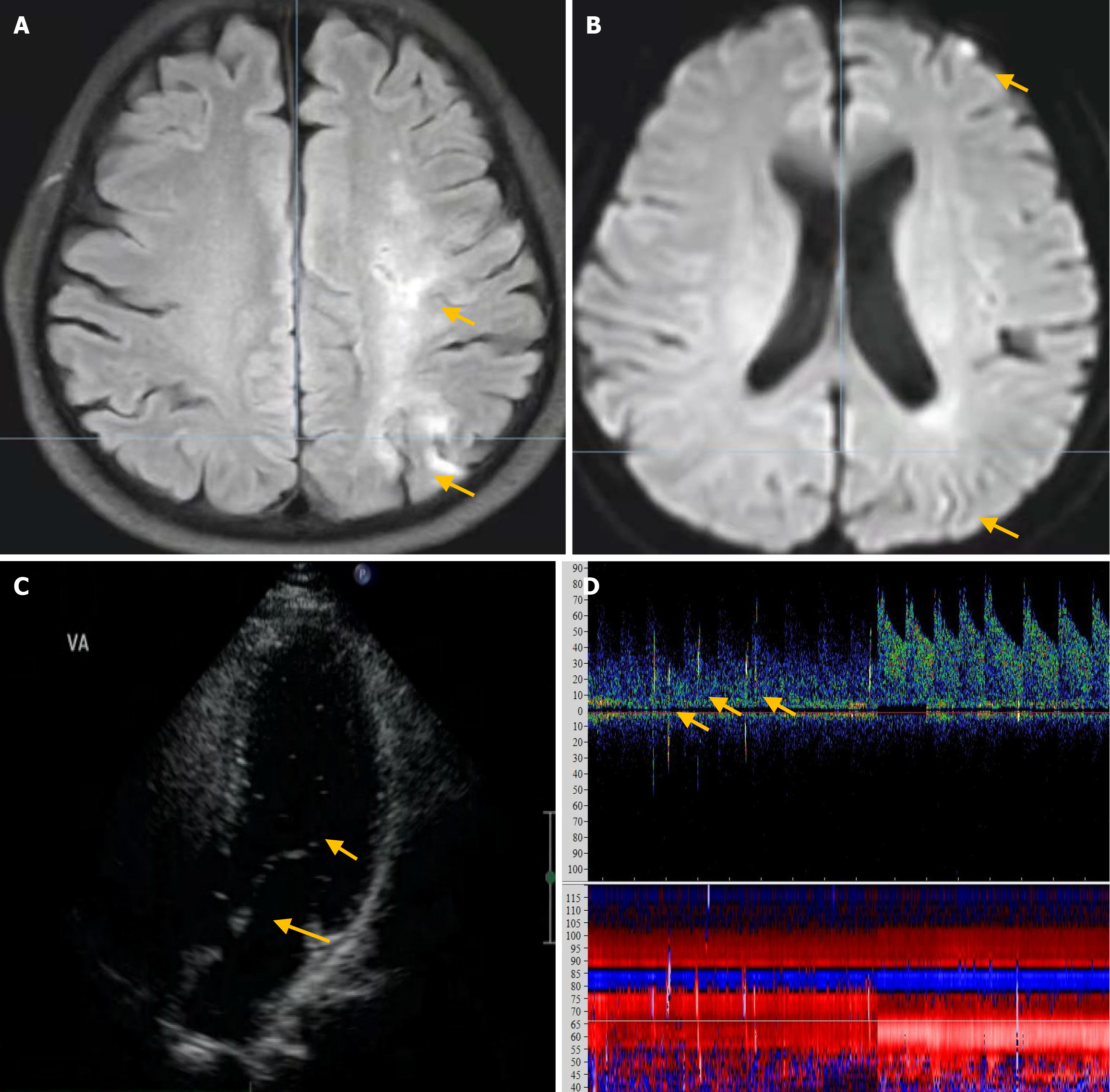
Figure 2 Brain magnetic resonance imaging revealing several infarcts located in the left occipital.
A: Arrow, fluid-attenuated inversion recovery sequences and frontal lobes; B: Arrow, diffusion-weighted imaging, as well as in the subcortical white matter; C: Contrast transthoracic echocardiography shows 11–30 microbubbles both at rest and during the Valsalva maneuver (VM); D: The contrast transcranial Doppler only detects approximately five microbubbles after the VM.
- Citation: Yao MJ, Zhao YY, Deng SP, Xiong HH, Wang J, Ren LJ, Cao LM. Right-to-left shunt detection via synchronized contrast transcranial Doppler combined with contrast transthoracic echocardiography: A preliminary study. World J Radiol 2024; 16(11): 657-667
- URL: https://www.wjgnet.com/1949-8470/full/v16/i11/657.htm
- DOI: https://dx.doi.org/10.4329/wjr.v16.i11.657


