Copyright
©The Author(s) 2024.
World J Radiol. Nov 28, 2024; 16(11): 644-656
Published online Nov 28, 2024. doi: 10.4329/wjr.v16.i11.644
Published online Nov 28, 2024. doi: 10.4329/wjr.v16.i11.644
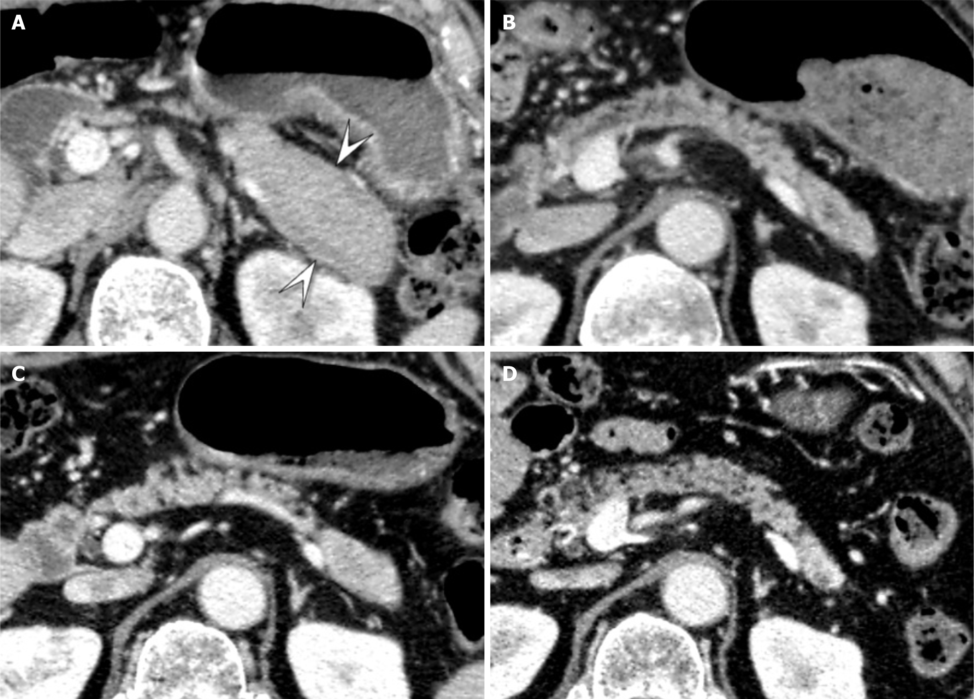
Figure 5 Portal venous contrast-enhanced computed tomography images of a 67-year-old man with mass-forming type autoimmune pancreatitis and re-enlargement during follow-up.
A: Initial computed tomography showing enlarged pancreatic tail with capsule-like rim (arrowheads); B: Six months later, pancreatic volume had markedly decreased (relative volume change 598%); C: Four years later, re-enlargement of the pancreatic tail with no capsule-like rim (arrowheads) was observed (relative volume change 683%); D: One year after re-enlargement, pancreatic volume had decreased again (relative volume change 589%).
- Citation: Shimada R, Yamada Y, Okamoto K, Murakami K, Motomura M, Takaki H, Fukuzawa K, Asayama Y. Pancreatic volume change using three dimensional-computed tomography volumetry and its relationships with diabetes on long-term follow-up in autoimmune pancreatitis. World J Radiol 2024; 16(11): 644-656
- URL: https://www.wjgnet.com/1949-8470/full/v16/i11/644.htm
- DOI: https://dx.doi.org/10.4329/wjr.v16.i11.644


