Copyright
©The Author(s) 2024.
World J Radiol. Nov 28, 2024; 16(11): 644-656
Published online Nov 28, 2024. doi: 10.4329/wjr.v16.i11.644
Published online Nov 28, 2024. doi: 10.4329/wjr.v16.i11.644
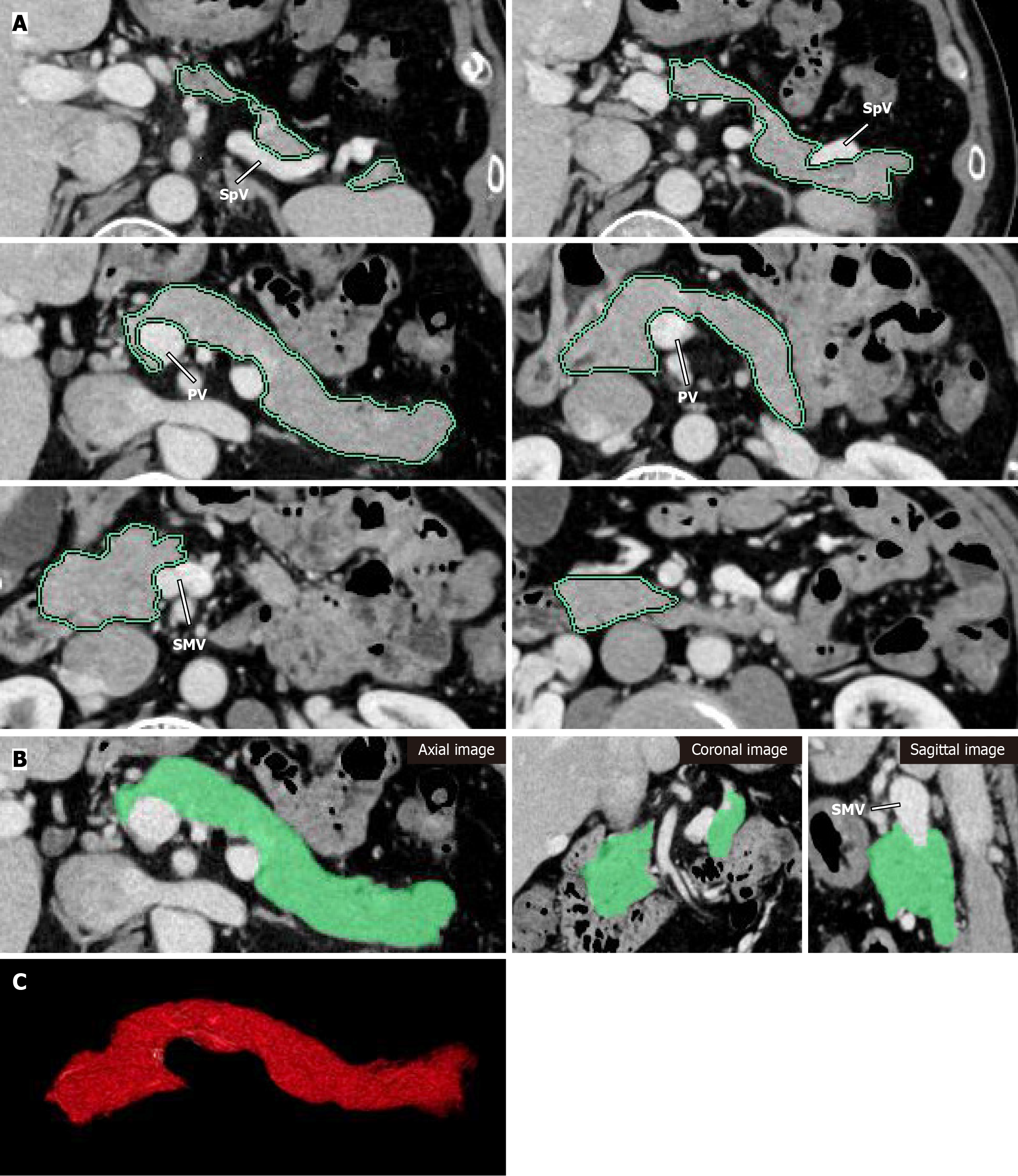
Figure 1 Pancreas volume measurement in a 62-year-old man with a normal pancreas, using portal venous contrast-enhanced computed tomography images.
A: Outline of pancreatic parenchyma was traced manually (green line) on each slice using a free region of interest (ROI) tool on contrast-enhanced-CT. While defining ROIs, care was taken to exclude the splenic artery and vein, superior mesenteric vein, and portal vein. The boundary of the pancreatic parenchyma was then outlined by extending the ROI using intensity-based semi-automated methods until the entire parenchyma in the slice was included; B: The traced pancreatic parenchyma was confirmed by the green area and manual correction was performed if misdelineation of the pancreatic borders was identified on axial, coronal, and sagittal images; C: The traced pancreatic parenchyma was transformed to the proximate shape in three dimensions and the pancreatic volume was then quantified. SpV: Splenic vein; PV: Portal vein; SMV: Superior mesenteric vein.
- Citation: Shimada R, Yamada Y, Okamoto K, Murakami K, Motomura M, Takaki H, Fukuzawa K, Asayama Y. Pancreatic volume change using three dimensional-computed tomography volumetry and its relationships with diabetes on long-term follow-up in autoimmune pancreatitis. World J Radiol 2024; 16(11): 644-656
- URL: https://www.wjgnet.com/1949-8470/full/v16/i11/644.htm
- DOI: https://dx.doi.org/10.4329/wjr.v16.i11.644


