Copyright
©The Author(s) 2024.
World J Radiol. Nov 28, 2024; 16(11): 629-637
Published online Nov 28, 2024. doi: 10.4329/wjr.v16.i11.629
Published online Nov 28, 2024. doi: 10.4329/wjr.v16.i11.629
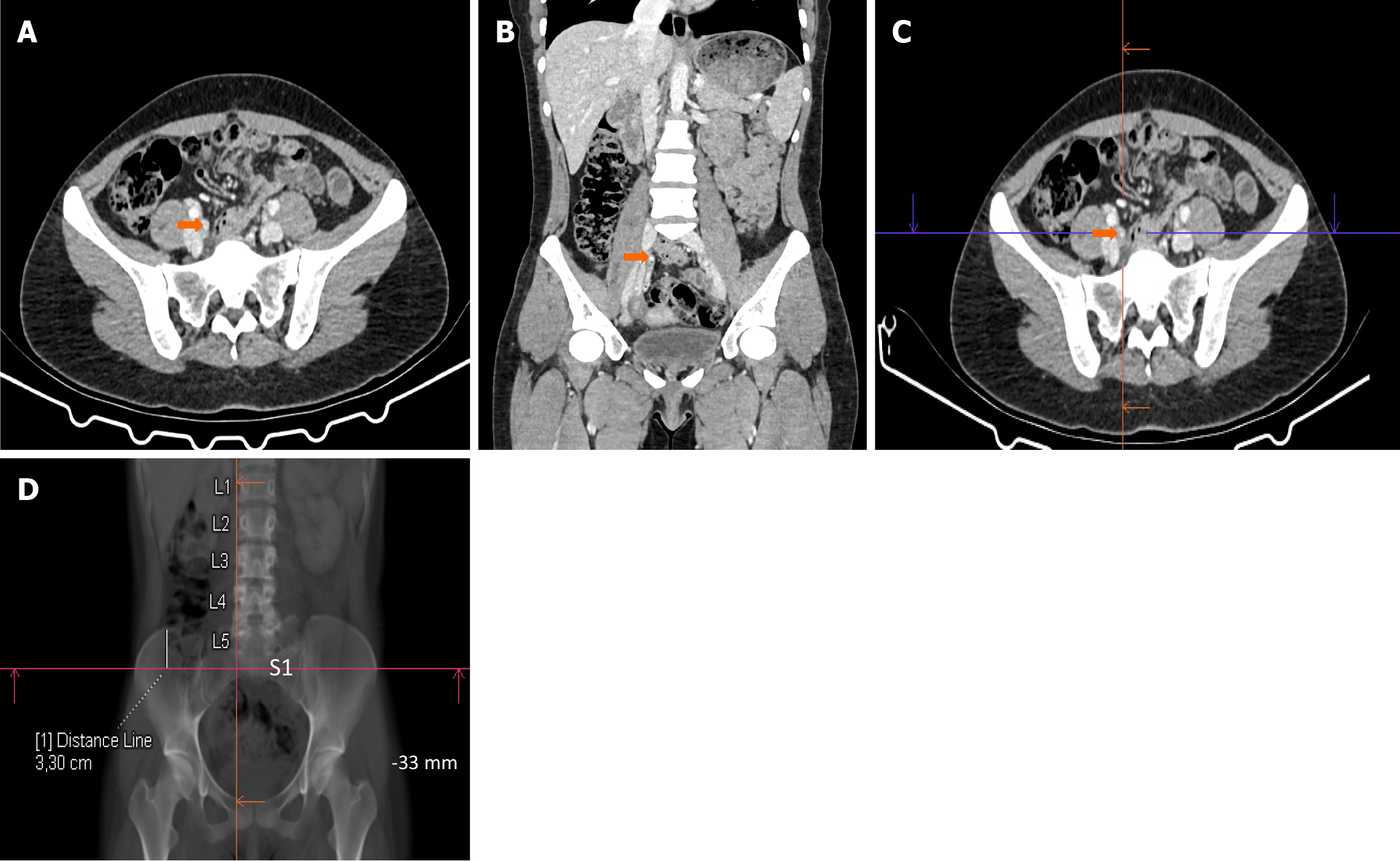
Figure 2 Determining the localization of the highest point of the appendix using the right iliac bone as a reference.
A: The highest point of the appendix vermiformis is detected (orange arrow) on axial images; B: The highest point is verified on coronal images (orange arrow); C: The highest point (orange arrow) is marked on axial images with reference lines; D: The distance between this designated point and the highest point of the right iliac bone is measured using the thick coronal multiplanar reconstruction images in the bone window. The distance is measured from the point where the horizontal line intersects with the highest point of the right iliac bone. If the designated point is situated above the highest point of the right iliac bone, it is denoted as positive (+), and if it is positioned below, it is denoted as negative (−), with measurements in mm. In this patient, the highest point of the appendix was localized at −33 mm relative to the highest point of the right iliac bone. Subsequently, using the same technique, the origin and lowest points of the appendix vermiformis were determined. L: Lumbar vertebra; S: Sacral vertebra.
- Citation: Ozturk MO, Resorlu M, Aydin S, Memis KB. Use of the vertebrae and iliac bone as references for localizing the appendix vermiformis in computed tomography. World J Radiol 2024; 16(11): 629-637
- URL: https://www.wjgnet.com/1949-8470/full/v16/i11/629.htm
- DOI: https://dx.doi.org/10.4329/wjr.v16.i11.629


