Copyright
©The Author(s) 2024.
World J Radiol. Oct 28, 2024; 16(10): 621-628
Published online Oct 28, 2024. doi: 10.4329/wjr.v16.i10.621
Published online Oct 28, 2024. doi: 10.4329/wjr.v16.i10.621
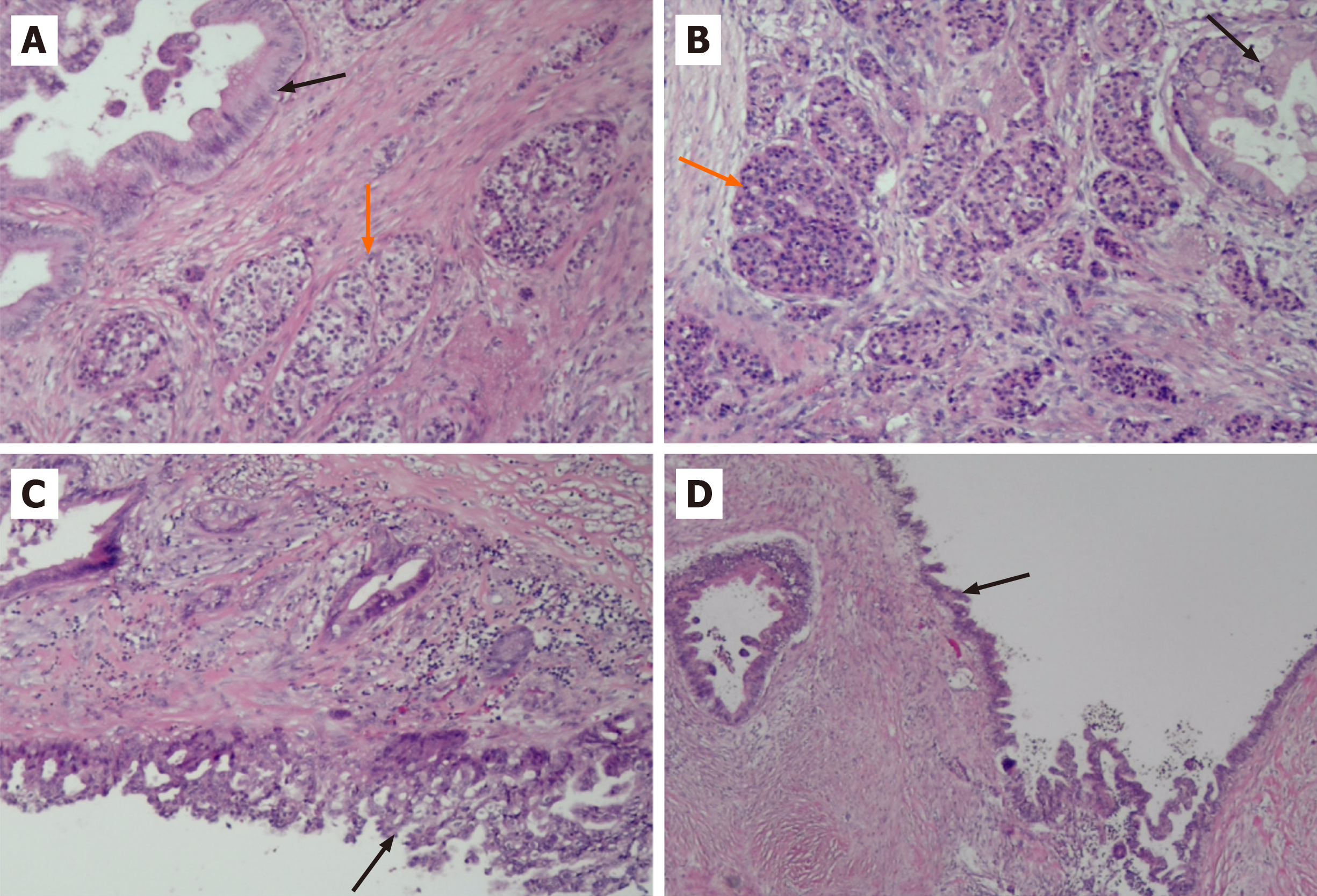
Figure 3 Ductal adenocarcinoma with neuroendocrine tumor.
A and B: The black arrow indicates ductal adenocarcinoma and the orange arrow indicates neuroendocrine tumor. H&E staining, magnification, ×100; C and D: The black arrow indicates the atypical epithelial cells lined with the cyst wall. H&E staining, magnification, x100.
- Citation: Zou DM, Shu ZY, Cao X. Cystic ductal adenocarcinoma of pancreas complicated with neuroendocrine tumor: A case report and review of literature. World J Radiol 2024; 16(10): 621-628
- URL: https://www.wjgnet.com/1949-8470/full/v16/i10/621.htm
- DOI: https://dx.doi.org/10.4329/wjr.v16.i10.621


