Copyright
©The Author(s) 2024.
World J Radiol. Oct 28, 2024; 16(10): 621-628
Published online Oct 28, 2024. doi: 10.4329/wjr.v16.i10.621
Published online Oct 28, 2024. doi: 10.4329/wjr.v16.i10.621
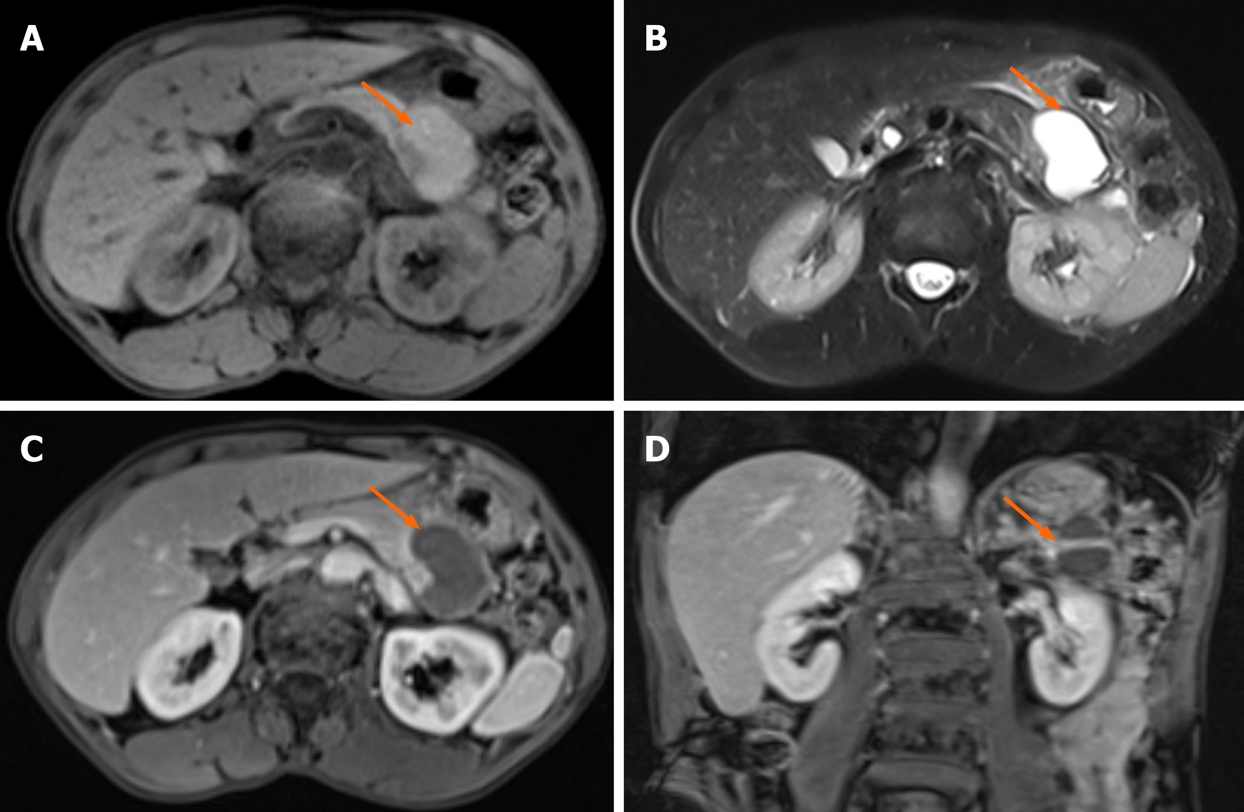
Figure 2 Preoperative magnetic resonance imaging results.
A: The cystic lesions (arrow) showed high signal intensity in the cyst and low signal intensity in the cyst wall on the T1-weighted axial images; B: The cystic lesions (arrow) showed obvious high signal intensity and the cystic wall showed low signal intensity on the T2-weighted axial images; C: Contrast-enhanced axial images showed enhancement of the cyst wall (arrow); D: The shape of the cyst (arrow) was irregular and the cyst wall was enhanced on the contrast-enhanced coronal images.
- Citation: Zou DM, Shu ZY, Cao X. Cystic ductal adenocarcinoma of pancreas complicated with neuroendocrine tumor: A case report and review of literature. World J Radiol 2024; 16(10): 621-628
- URL: https://www.wjgnet.com/1949-8470/full/v16/i10/621.htm
- DOI: https://dx.doi.org/10.4329/wjr.v16.i10.621


