Copyright
©The Author(s) 2024.
World J Radiol. Oct 28, 2024; 16(10): 608-615
Published online Oct 28, 2024. doi: 10.4329/wjr.v16.i10.608
Published online Oct 28, 2024. doi: 10.4329/wjr.v16.i10.608
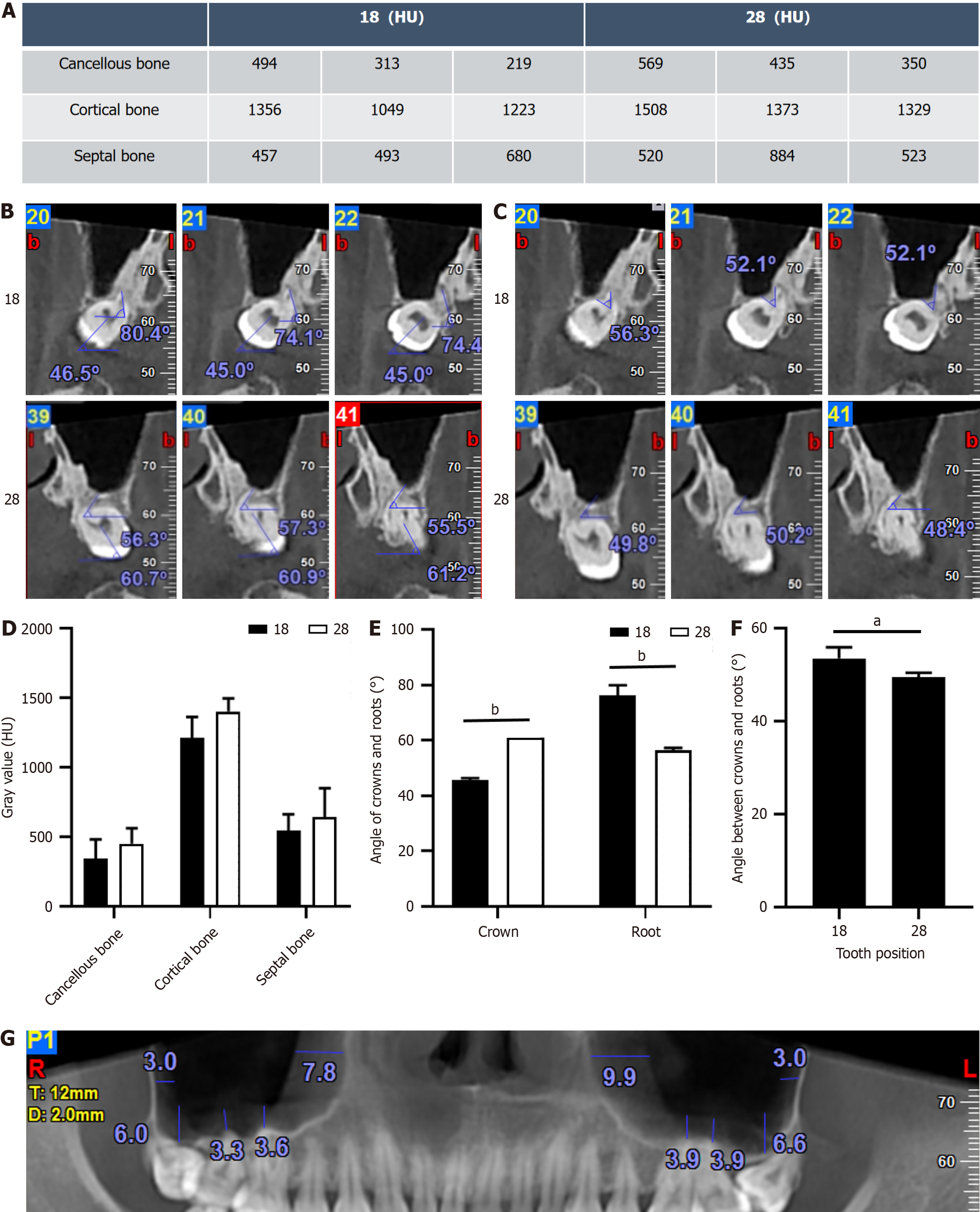
Figure 5 Digital imaging analysis of secondary factors in cone-beam computed tomography.
A: Measurement of gray values; B: Angle measurement between the crown and root relative to the jaw plane; C: Angle measurement between crowns and roots; D: Statistical analysis of gray values at different positions; E: Statistical analysis of the angle between the crown and root relative to the Frankfort plane; F: Statistical analysis of the angle between crowns and roots; G: Measurement of maxillary sinus mucosa. aP < 0.05, bP < 0.001.
- Citation: Liu H, Wang F, Tang YL, Yan X. Asymmetric outcomes in bilateral maxillary impacted tooth extractions: A case report. World J Radiol 2024; 16(10): 608-615
- URL: https://www.wjgnet.com/1949-8470/full/v16/i10/608.htm
- DOI: https://dx.doi.org/10.4329/wjr.v16.i10.608


