Copyright
©The Author(s) 2024.
World J Radiol. Oct 28, 2024; 16(10): 608-615
Published online Oct 28, 2024. doi: 10.4329/wjr.v16.i10.608
Published online Oct 28, 2024. doi: 10.4329/wjr.v16.i10.608
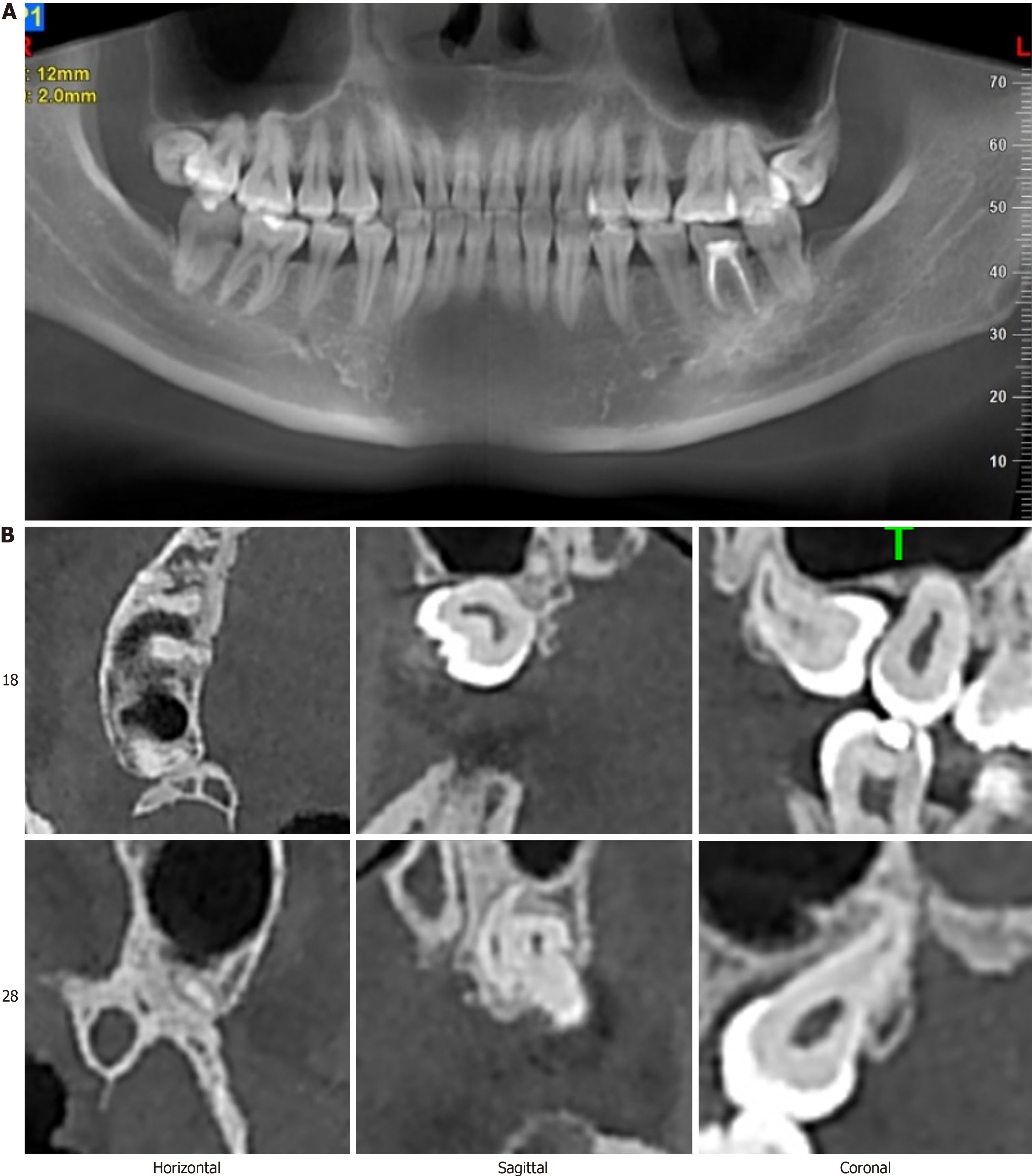
Figure 2 Preoperative imaging.
A: Surface slice; B: Cone-beam computed tomography images at three levels. In the sagittal view, a portion of the crown of tooth 18 is in contact with the maxillary sinus, while in both sagittal and coronal views, part of the root of tooth 28 is seen to abut the maxillary sinus.
- Citation: Liu H, Wang F, Tang YL, Yan X. Asymmetric outcomes in bilateral maxillary impacted tooth extractions: A case report. World J Radiol 2024; 16(10): 608-615
- URL: https://www.wjgnet.com/1949-8470/full/v16/i10/608.htm
- DOI: https://dx.doi.org/10.4329/wjr.v16.i10.608


