Copyright
©The Author(s) 2024.
World J Radiol. Oct 28, 2024; 16(10): 593-599
Published online Oct 28, 2024. doi: 10.4329/wjr.v16.i10.593
Published online Oct 28, 2024. doi: 10.4329/wjr.v16.i10.593
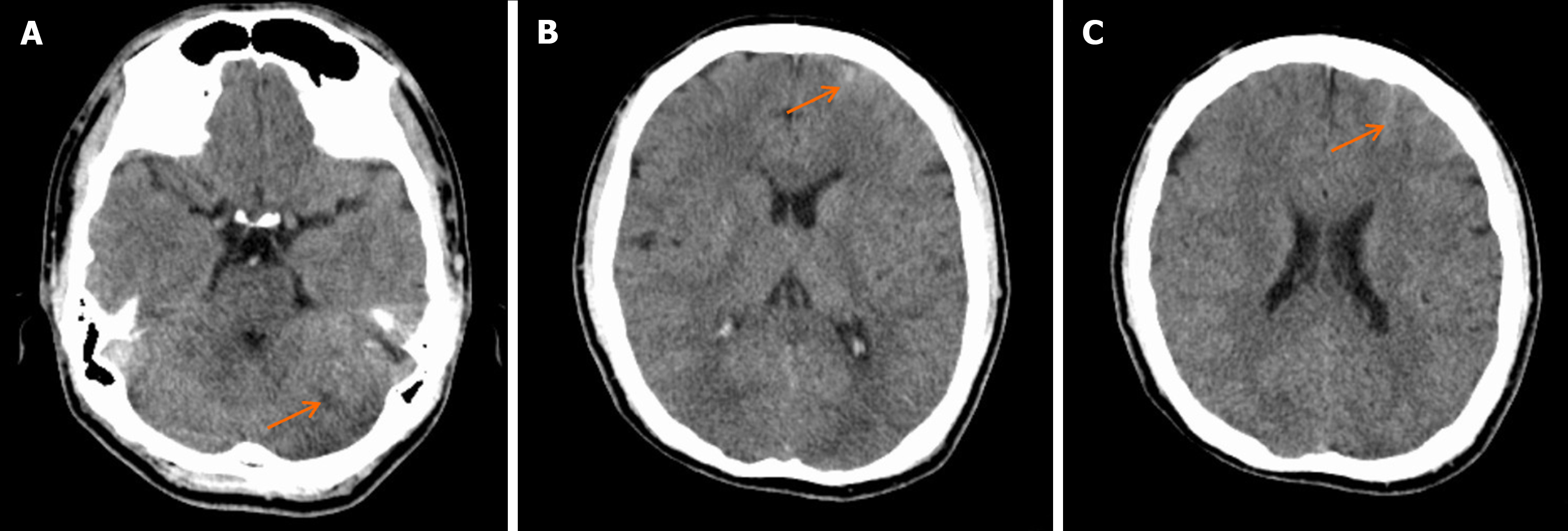
Figure 1 Head computed tomography image upon admission.
A: Bilateral cerebellar hemispheres show signs of suspicious ischemia; B: Possible hematoma in the left frontal lobe; C: Left frontal sulci subarachnoid hemorrhage.
- Citation: Zhang HB, Duan YH, Zhou M, Liang RC. High-resolution magnetic resonance imaging in the diagnosis and management of vertebral artery dissection: A case report. World J Radiol 2024; 16(10): 593-599
- URL: https://www.wjgnet.com/1949-8470/full/v16/i10/593.htm
- DOI: https://dx.doi.org/10.4329/wjr.v16.i10.593


