Copyright
©The Author(s) 2024.
World J Radiol. Oct 28, 2024; 16(10): 586-592
Published online Oct 28, 2024. doi: 10.4329/wjr.v16.i10.586
Published online Oct 28, 2024. doi: 10.4329/wjr.v16.i10.586
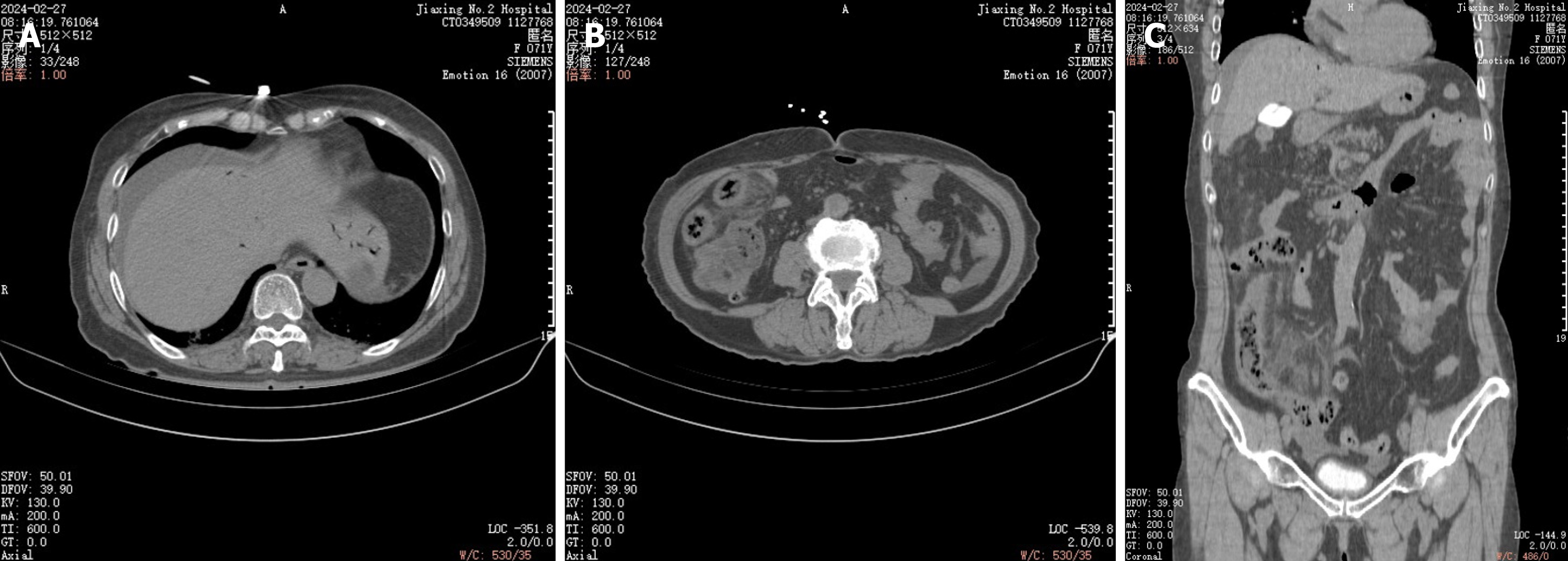
Figure 4 Comparison of computerized tomography scan on February 26, 2024, with necrosis of the bowel wall, peripheral exudative accumulation of mesenteric edema progressing more than before, and additional abdominopelvic effusion.
Superior mesenteric vein and its collateral vessels with multiple pneumoperitoneum have been absorbed compared to before, and intrahepatic portal vein pneumoperitoneum has been significantly absorbed compared to before. A: Transverse section of the liver; B: Transverse section of the intestinal lumen; C: Sagittal view.
- Citation: Yu ZX, Bin Z, Lun ZK, Jiang XJ. Portal venous gas complication following coronary angiography: A case report. World J Radiol 2024; 16(10): 586-592
- URL: https://www.wjgnet.com/1949-8470/full/v16/i10/586.htm
- DOI: https://dx.doi.org/10.4329/wjr.v16.i10.586


