Copyright
©The Author(s) 2024.
World J Radiol. Oct 28, 2024; 16(10): 586-592
Published online Oct 28, 2024. doi: 10.4329/wjr.v16.i10.586
Published online Oct 28, 2024. doi: 10.4329/wjr.v16.i10.586
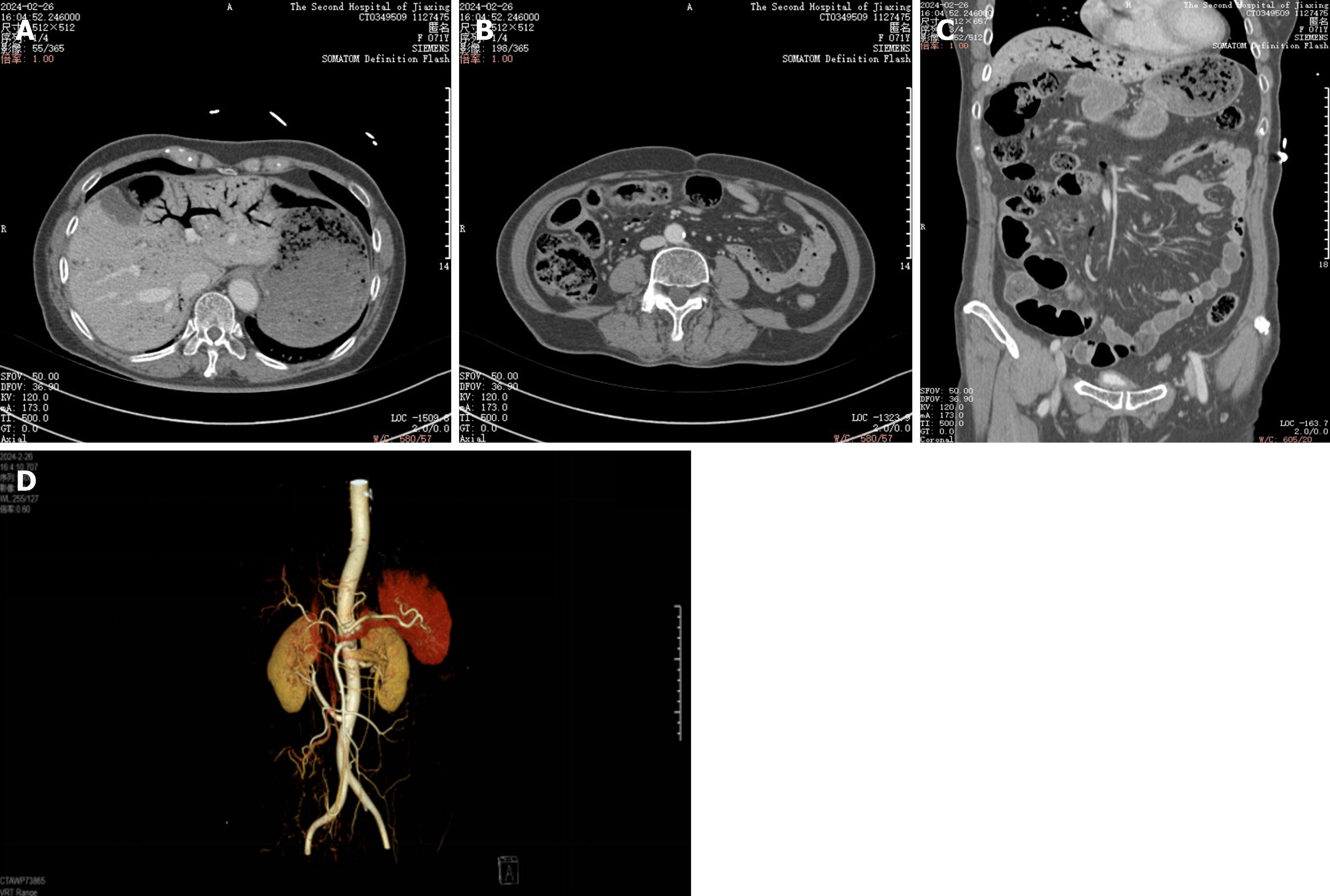
Figure 3 Emergency computerized tomography scan of abdominal aortic enhancement.
A: Transverse section of the liver; B: Transverse section of the intestinal lumen; C: Sagittal view. A-C: Extensive pneumoperitoneum in the portal vein. Multiple scattered gas in the superior mesenteric vein and its collateral branches (right middle and lower abdominal peristomal vessels), turbid spaces of the right middle and lower abdominal peristomal fat, and a localised small bowel wall in the right lower abdomen; D: Reconstruction of the vascular image of the abdominal aorta: Localised calcified plaque in the abdominal aorta, compression syndrome of the median arcuate ligament, no embolism of the mesenteric artery.
- Citation: Yu ZX, Bin Z, Lun ZK, Jiang XJ. Portal venous gas complication following coronary angiography: A case report. World J Radiol 2024; 16(10): 586-592
- URL: https://www.wjgnet.com/1949-8470/full/v16/i10/586.htm
- DOI: https://dx.doi.org/10.4329/wjr.v16.i10.586


