Copyright
©The Author(s) 2024.
World J Radiol. Oct 28, 2024; 16(10): 569-578
Published online Oct 28, 2024. doi: 10.4329/wjr.v16.i10.569
Published online Oct 28, 2024. doi: 10.4329/wjr.v16.i10.569
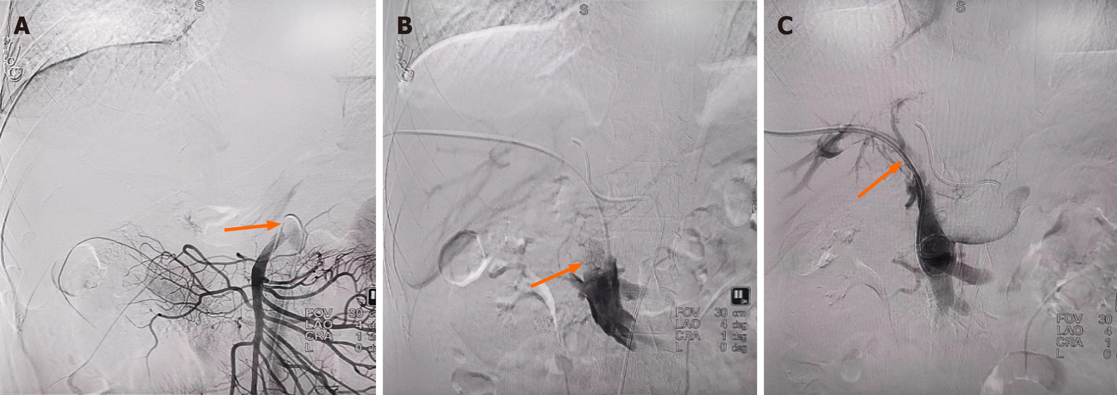
Figure 2 Indirect and direct portal venography conducted on February 29, 2024.
A: Indirect portal venography showed an underdeveloped portal vein (orange arrows); B: Direct portal vein angiography showed significant filling defects (orange arrows), indicating thrombosis; C: Partial visualisation of the portal vein (orange arrows) during intraoperative angiography after thrombolysis via an indwelling biliary drainage tube.
- Citation: Yuan JJ, Zhang HF, Zhang J, Li JZ. Mesenteric venous thrombosis in a young adult: A case report and review of the literature. World J Radiol 2024; 16(10): 569-578
- URL: https://www.wjgnet.com/1949-8470/full/v16/i10/569.htm
- DOI: https://dx.doi.org/10.4329/wjr.v16.i10.569


