Copyright
©The Author(s) 2024.
World J Radiol. Oct 28, 2024; 16(10): 537-544
Published online Oct 28, 2024. doi: 10.4329/wjr.v16.i10.537
Published online Oct 28, 2024. doi: 10.4329/wjr.v16.i10.537
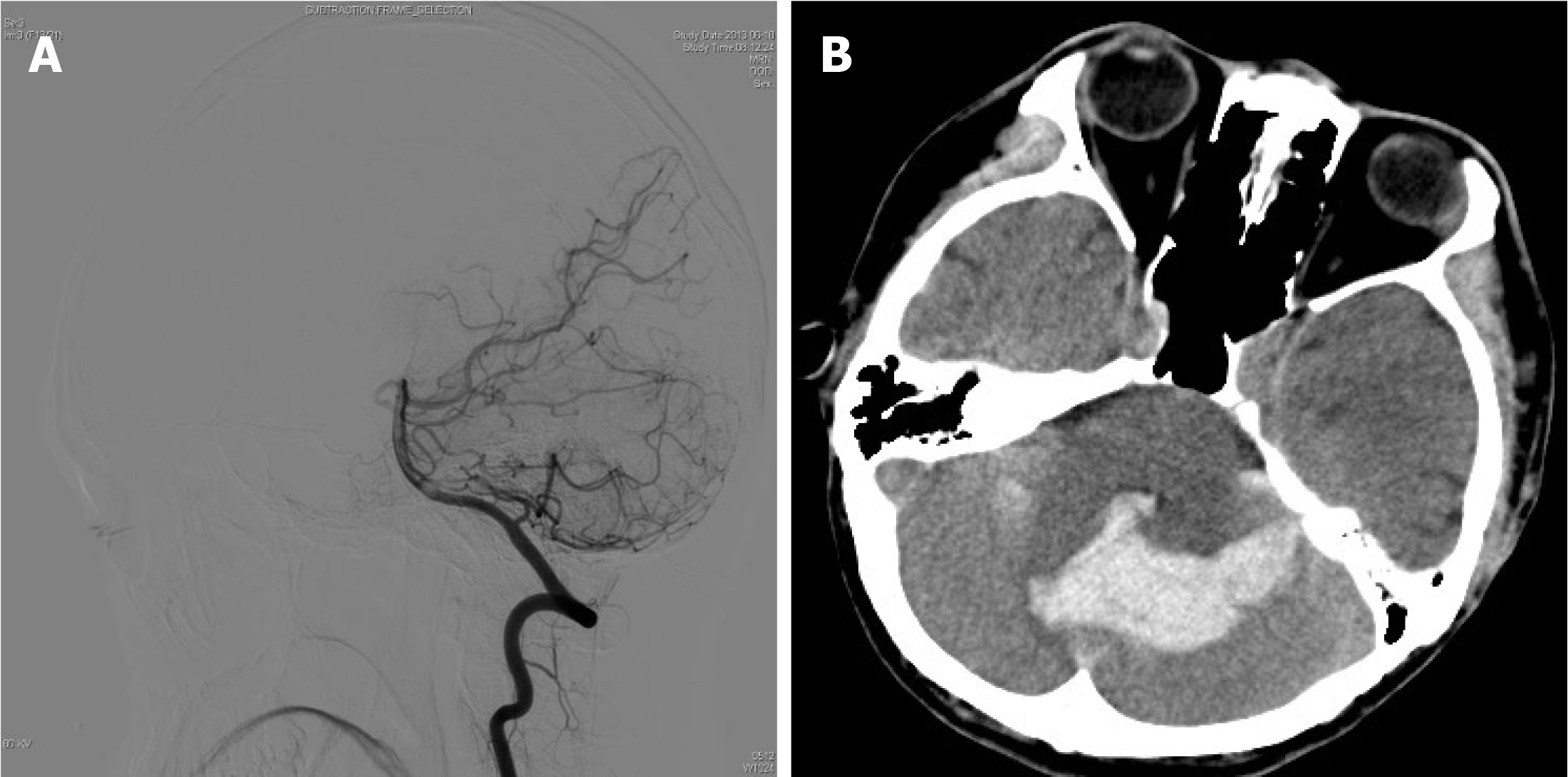
Figure 3 Right vertebral artery angiography and Preoperative computed tomography.
A: Right vertebral artery angiography performed before the first arteriovenous malformation resection showing no cerebellar abnormalities; B: Preoperative computed tomography showing multiple patchy hyperdense shadows in the 3rd and 4th ventricles, indicating bilateral cerebellar hemorrhage.
- Citation: Cao WY, Li JP, Guo P, Song LX. Ectopic recurrence following treatment of arteriovenous malformations in an adult: A case report and review of literature. World J Radiol 2024; 16(10): 537-544
- URL: https://www.wjgnet.com/1949-8470/full/v16/i10/537.htm
- DOI: https://dx.doi.org/10.4329/wjr.v16.i10.537


