Copyright
©The Author(s) 2024.
World J Radiol. Oct 28, 2024; 16(10): 528-536
Published online Oct 28, 2024. doi: 10.4329/wjr.v16.i10.528
Published online Oct 28, 2024. doi: 10.4329/wjr.v16.i10.528
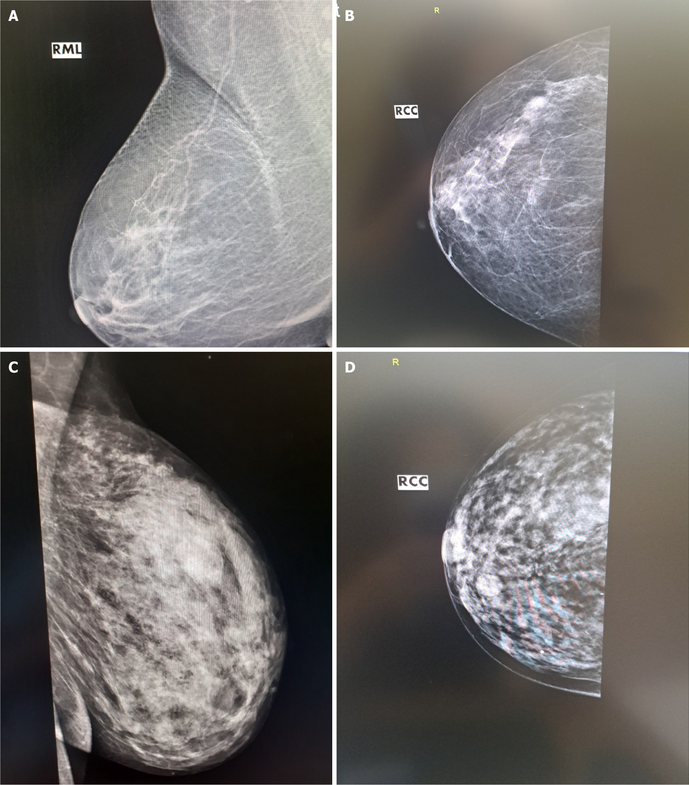
Figure 2 Mammography in mediolateral oblique and craniocaudal view.
A: Ridge-like lesion in the upper lateral quadrant is shown, which is classified as Breast Imaging Reporting and Data System 5; B: In the right mammary gland in the upper lateral quadrant, a dense, spiculated lesion is visualized. The lesion has very high suspicion of malignant tumor and is classified as Breast Imaging Reporting and Data System 5; C: In the right mammary gland, a thickened area is palpated in the upper lateral quadrant (the most common location of breast carcinoma). Mammographically, the gland is presented with a dense structure and has low informativeness. However, an area of further increased density is seen in the palpably thickened area; D: In the right mammary gland, in the lower medial quadrant, a rounded strong shadow with almost smooth outlines is visualized. Ventrally, the cyst-like finding has an uneven contour. This uneven contour necessitates sonographic refinement of the lesion. RCC: Right craniocaudal; RML: Right mediolateral.
- Citation: Chervenkov L, Georgiev A, Doykov M, Velikova T. Breast cancer imaging-clinical experience with two-dimensional-shear wave elastography: A retrospective study. World J Radiol 2024; 16(10): 528-536
- URL: https://www.wjgnet.com/1949-8470/full/v16/i10/528.htm
- DOI: https://dx.doi.org/10.4329/wjr.v16.i10.528


