Copyright
©The Author(s) 2024.
World J Radiol. Oct 28, 2024; 16(10): 528-536
Published online Oct 28, 2024. doi: 10.4329/wjr.v16.i10.528
Published online Oct 28, 2024. doi: 10.4329/wjr.v16.i10.528
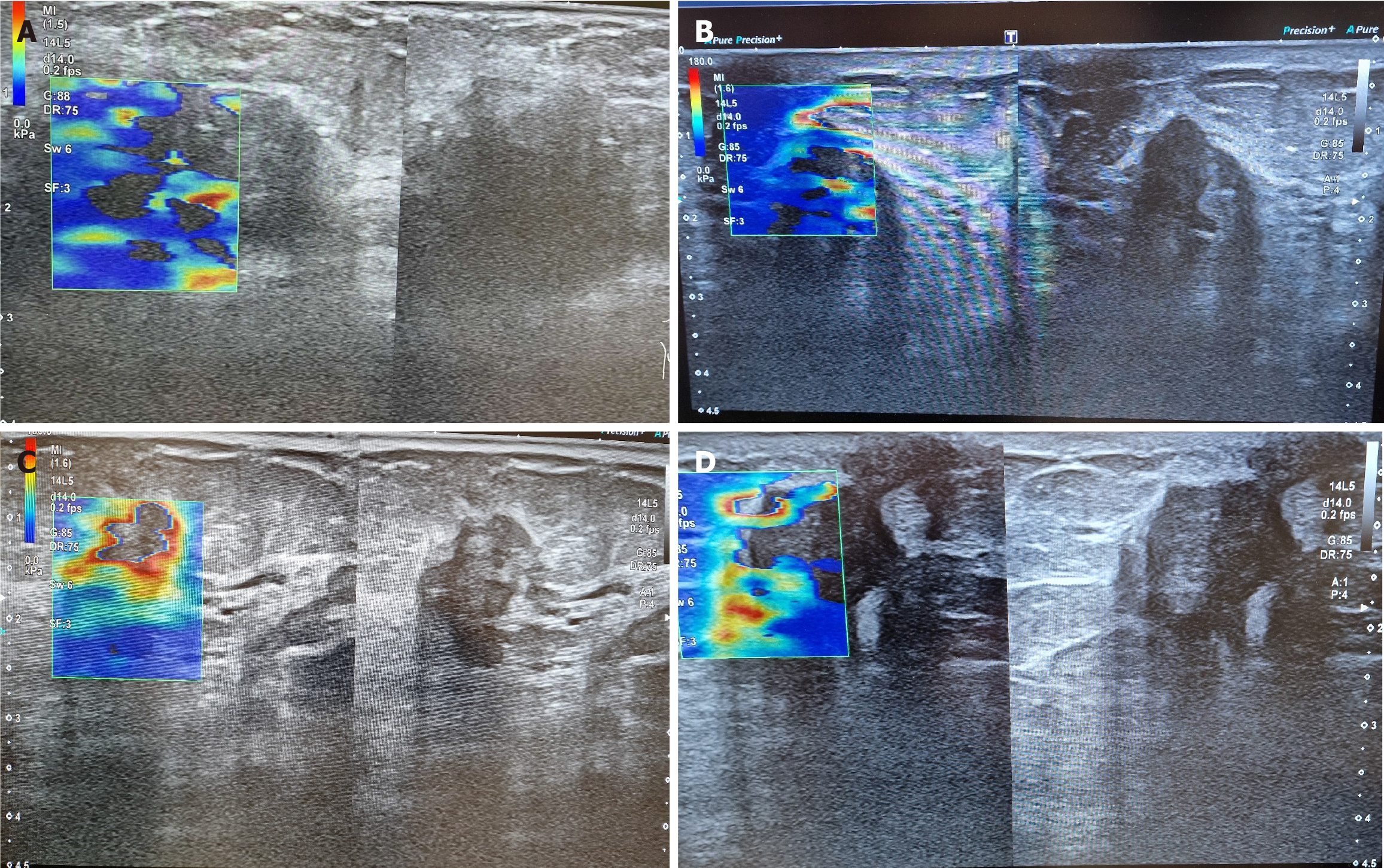
Figure 1 Two-dimensional-shear wave elastography.
A: A solid malignant lesion in the upper medial quadrant of the right mammary gland is presented. The finding has an irregular (ovoid) shape and is oriented antiparallel to the glandular parenchyma. The lesion has been scored Breast Imaging Reporting and Data System 5 tumor. Elastography demonstrated increased peripheral stiffness. After biopsy, the finding proved to be invasive ductal carcinoma; B: Formation with irregular form was found, which is inhomogeneous and is orientated antiparallel to the parenchyma. The lesion is bigger in height than in width. Elastography showed non-uniform areas of increased density. The finding was determined as Breast Imaging Reporting and Data System category 5 and was confirmed as malignant by a fine-needle biopsy; C: Solid formation with irregular form is found, which is antiparallel to the parenchyma. On elastographic evaluation, we see a peripherally increased eggshell-like density. This type of increased peripheral density is due to desmoplasia. The tumor is staged Breast Imaging Reporting and Data System 5; D: Formation, previously described as a cyst on mammography is found on this examination. The tumor is solid and has no characteristics of cystic lesion. Elastographically, non-uniform density zones in the find are also distinguished. The find is scored as Breast Imaging Reporting and Data System 4C, fine needle biopsy confirmed ductal invasive carcinoma.
- Citation: Chervenkov L, Georgiev A, Doykov M, Velikova T. Breast cancer imaging-clinical experience with two-dimensional-shear wave elastography: A retrospective study. World J Radiol 2024; 16(10): 528-536
- URL: https://www.wjgnet.com/1949-8470/full/v16/i10/528.htm
- DOI: https://dx.doi.org/10.4329/wjr.v16.i10.528


