Copyright
©The Author(s) 2024.
World J Radiol. Oct 28, 2024; 16(10): 497-511
Published online Oct 28, 2024. doi: 10.4329/wjr.v16.i10.497
Published online Oct 28, 2024. doi: 10.4329/wjr.v16.i10.497
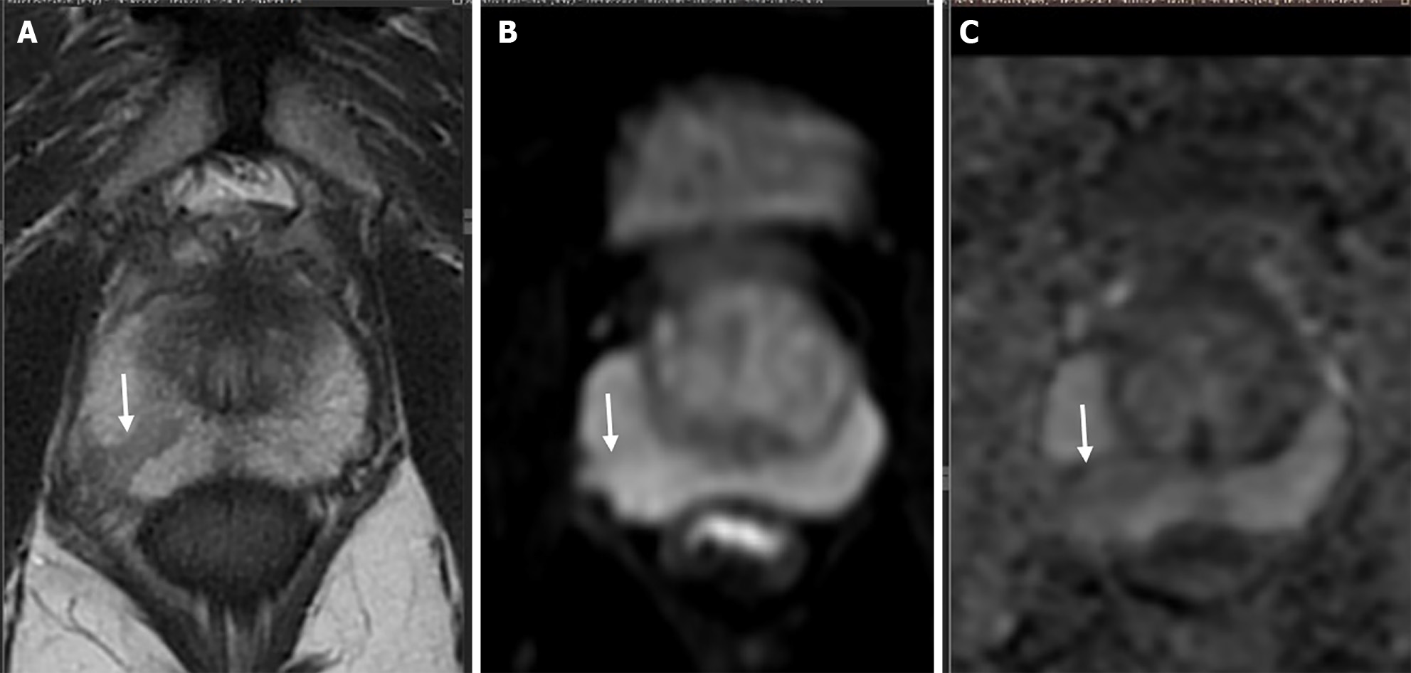
Figure 3 Shape analysis.
A-C: T2-weighted axial image shows a wedge-shaped T2 hypointense lesion (arrow in A) in right peripheral zone which shows no diffusion restriction (arrow in B and C) is scored as a category 2 observation according to PIRADS v.2.1.
- Citation: Dhiman A, Kumar V, Das CJ. Quantitative magnetic resonance imaging in prostate cancer: A review of current technology. World J Radiol 2024; 16(10): 497-511
- URL: https://www.wjgnet.com/1949-8470/full/v16/i10/497.htm
- DOI: https://dx.doi.org/10.4329/wjr.v16.i10.497


