Copyright
©The Author(s) 2023.
World J Radiol. Sep 28, 2023; 15(9): 274-280
Published online Sep 28, 2023. doi: 10.4329/wjr.v15.i9.274
Published online Sep 28, 2023. doi: 10.4329/wjr.v15.i9.274
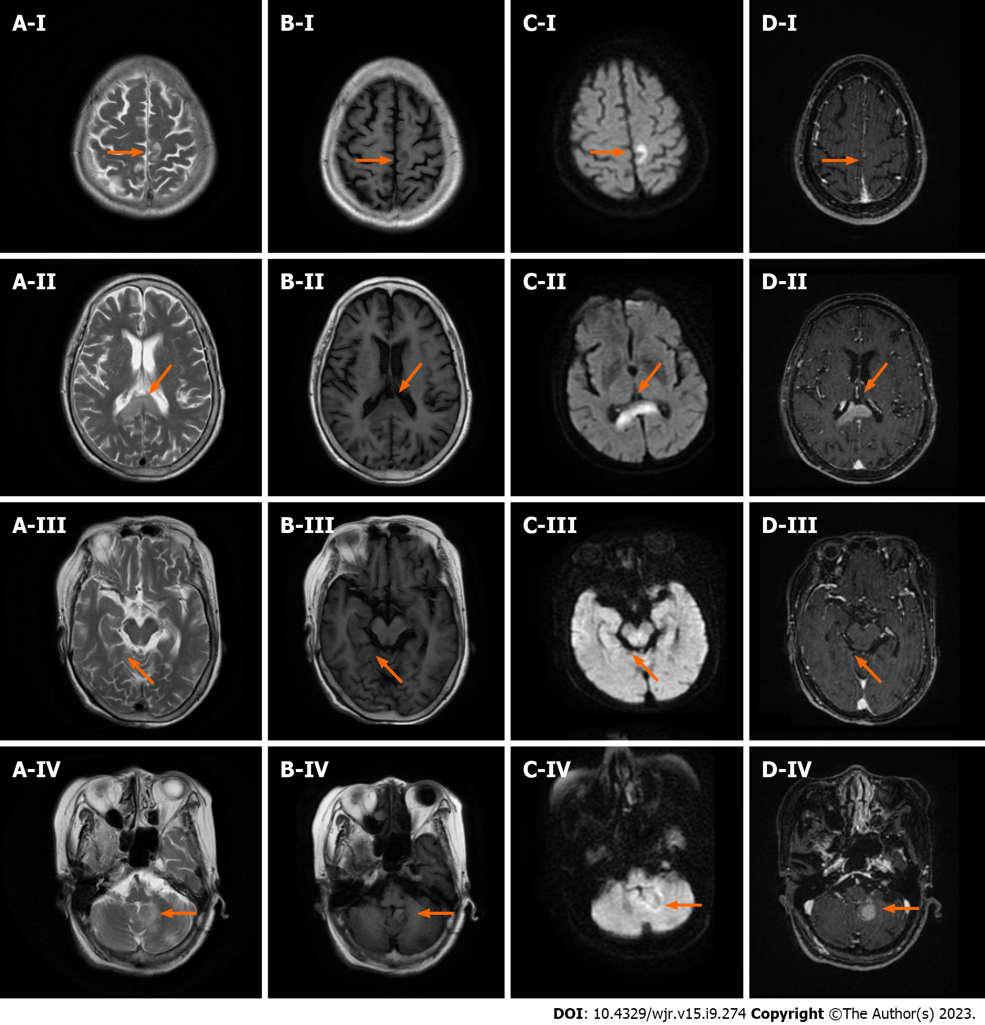
Figure 3 Images of the patient following corticosteroid pulse therapy.
A: T2WI; B: T1WI; C: Diffusion-weighted imaging; D: T1-weighted enhanced scan; I: Parietal lobe lesions; II: Corpus callosum lesions; III: Hippocampal lesions; IV: Left cerebellar hemisphere lesions.
- Citation: Liu LH, Zhang HW, Zhang HB, Liu XL, Deng HZ, Lin F, Huang B. Distinctive magnetic resonance imaging features in primary central nervous system lymphoma: A case report. World J Radiol 2023; 15(9): 274-280
- URL: https://www.wjgnet.com/1949-8470/full/v15/i9/274.htm
- DOI: https://dx.doi.org/10.4329/wjr.v15.i9.274


