Copyright
©The Author(s) 2023.
World J Radiol. Sep 28, 2023; 15(9): 256-273
Published online Sep 28, 2023. doi: 10.4329/wjr.v15.i9.256
Published online Sep 28, 2023. doi: 10.4329/wjr.v15.i9.256
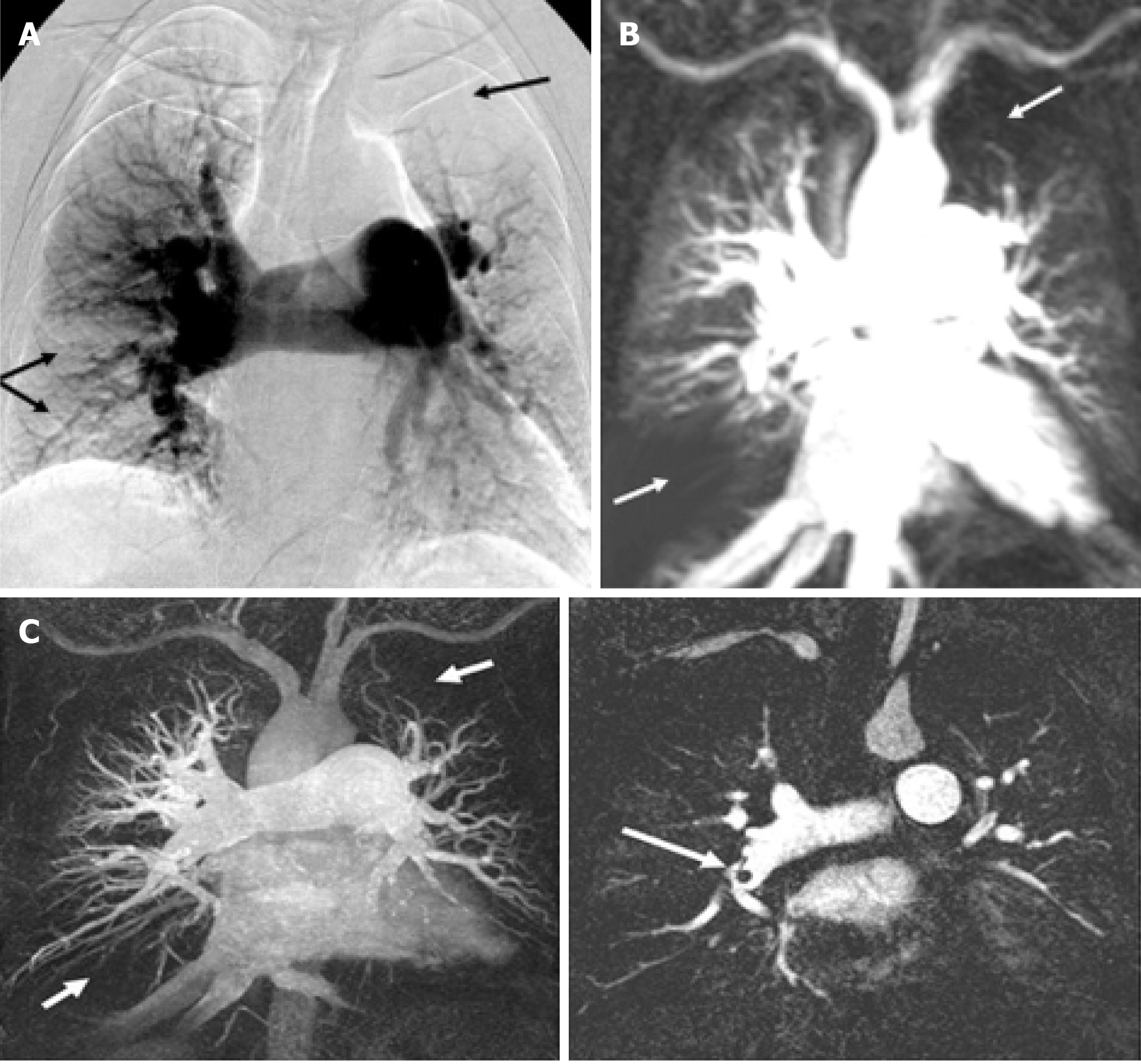
Figure 2 Comparing digital subtraction angiography, pulmonary perfusion magnetic resonance imaging and magnetic resonance angiography for identifying perfusion defects in chronic thromboembolic pulmonary hypertension patient.
A: Digital subtraction angiography showing perfusion defects (arrows) in left upper lobe and right lower lobe due to thromboembolic occlusion; B: Magnetic resonance imaging perfusion image at peak enhancement showing hypo-intense perfusion defects at same locations; C: Magnetic resonance angiogram of the same patient demonstrating dark thromboembolic material in the right pulmonary artery corresponding to perfusion defects at the same location. Citation: Nikolaou K, Schoenberg SO, Attenberger U, Scheidler J, Dietrich O, Kuehn B, Rosa F, Huber A, Leuchte H, Baumgartner R, Behr J, Reiser MF. Pulmonary arterial hypertension: diagnosis with fast perfusion MR imaging and high-spatial-resolution MR angiography--preliminary experience. Radiology 2005; 236: 694-703 [PMID: 15994997 DOI: 10.1148/radiol.2361040502].
- Citation: Lacharie M, Villa A, Milidonis X, Hasaneen H, Chiribiri A, Benedetti G. Role of pulmonary perfusion magnetic resonance imaging for the diagnosis of pulmonary hypertension: A review. World J Radiol 2023; 15(9): 256-273
- URL: https://www.wjgnet.com/1949-8470/full/v15/i9/256.htm
- DOI: https://dx.doi.org/10.4329/wjr.v15.i9.256


