Copyright
©The Author(s) 2023.
World J Radiol. Jul 28, 2023; 15(7): 234-240
Published online Jul 28, 2023. doi: 10.4329/wjr.v15.i7.234
Published online Jul 28, 2023. doi: 10.4329/wjr.v15.i7.234
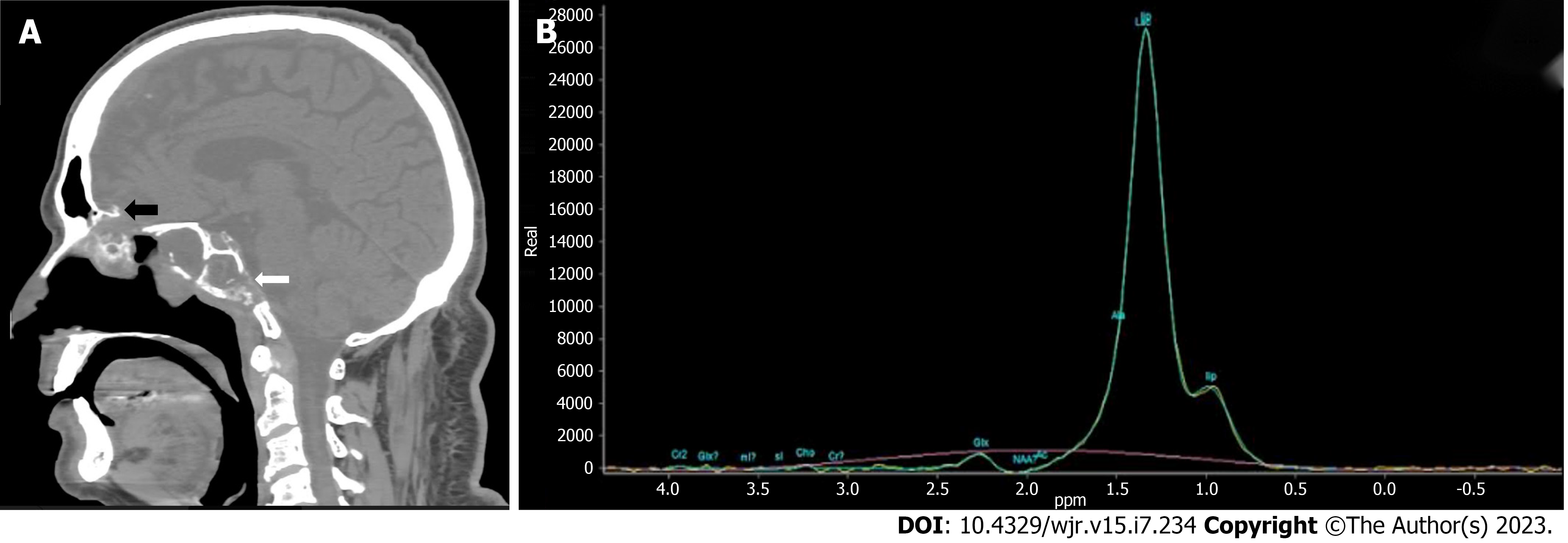
Figure 4 Images of patient with post COVID-19 mucormycosis.
A: Computerized tomography of brain after one month mid sagittal reformatted section in bone window demonstrating destruction of cribriform plate (black arrow), sphenoid sinus and clivus (white arrow); B: Magnetic resonance spectroscopy with a voxel placed in right frontal lobe fungal abscess demonstrating lactate peak at 1.33 ppm.
- Citation: Narra R, Rayapati S. Invasive rhinocerebral mucormycosis: Imaging the temporal evolution of disease in post COVID-19 case with diabetes: A case report. World J Radiol 2023; 15(7): 234-240
- URL: https://www.wjgnet.com/1949-8470/full/v15/i7/234.htm
- DOI: https://dx.doi.org/10.4329/wjr.v15.i7.234


