Copyright
©The Author(s) 2023.
World J Radiol. Jul 28, 2023; 15(7): 234-240
Published online Jul 28, 2023. doi: 10.4329/wjr.v15.i7.234
Published online Jul 28, 2023. doi: 10.4329/wjr.v15.i7.234
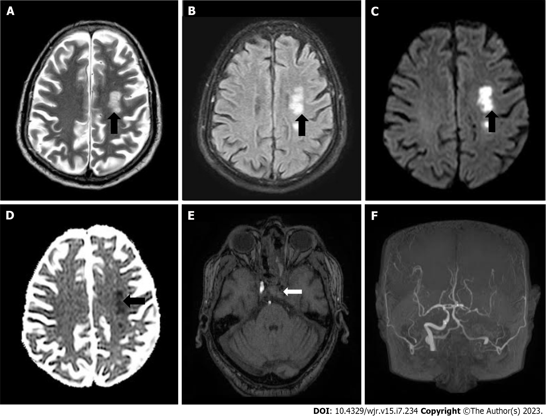
Figure 2 Images of patient with post COVID-19 mucormycosis.
A and B: Axial T2-weighted and axial fluid attenuated inversion recovery images of brain 15 d post admission demonstrating acute infarcts (black arrows); C and D: In left centrum semiovale with diffusion restriction on diffusion-weighted imaging and reversal on apparent diffusion coefficient represented by black arrows; E and F: Magnetic resonance angiography (MRA) images of Brain demonstrating loss of flow signal is noted in left internal carotid artery (white arrow) on source images and absent left internal carotid artery on maximum intensity projection MRA.
- Citation: Narra R, Rayapati S. Invasive rhinocerebral mucormycosis: Imaging the temporal evolution of disease in post COVID-19 case with diabetes: A case report. World J Radiol 2023; 15(7): 234-240
- URL: https://www.wjgnet.com/1949-8470/full/v15/i7/234.htm
- DOI: https://dx.doi.org/10.4329/wjr.v15.i7.234


