Copyright
©The Author(s) 2023.
World J Radiol. Jul 28, 2023; 15(7): 234-240
Published online Jul 28, 2023. doi: 10.4329/wjr.v15.i7.234
Published online Jul 28, 2023. doi: 10.4329/wjr.v15.i7.234
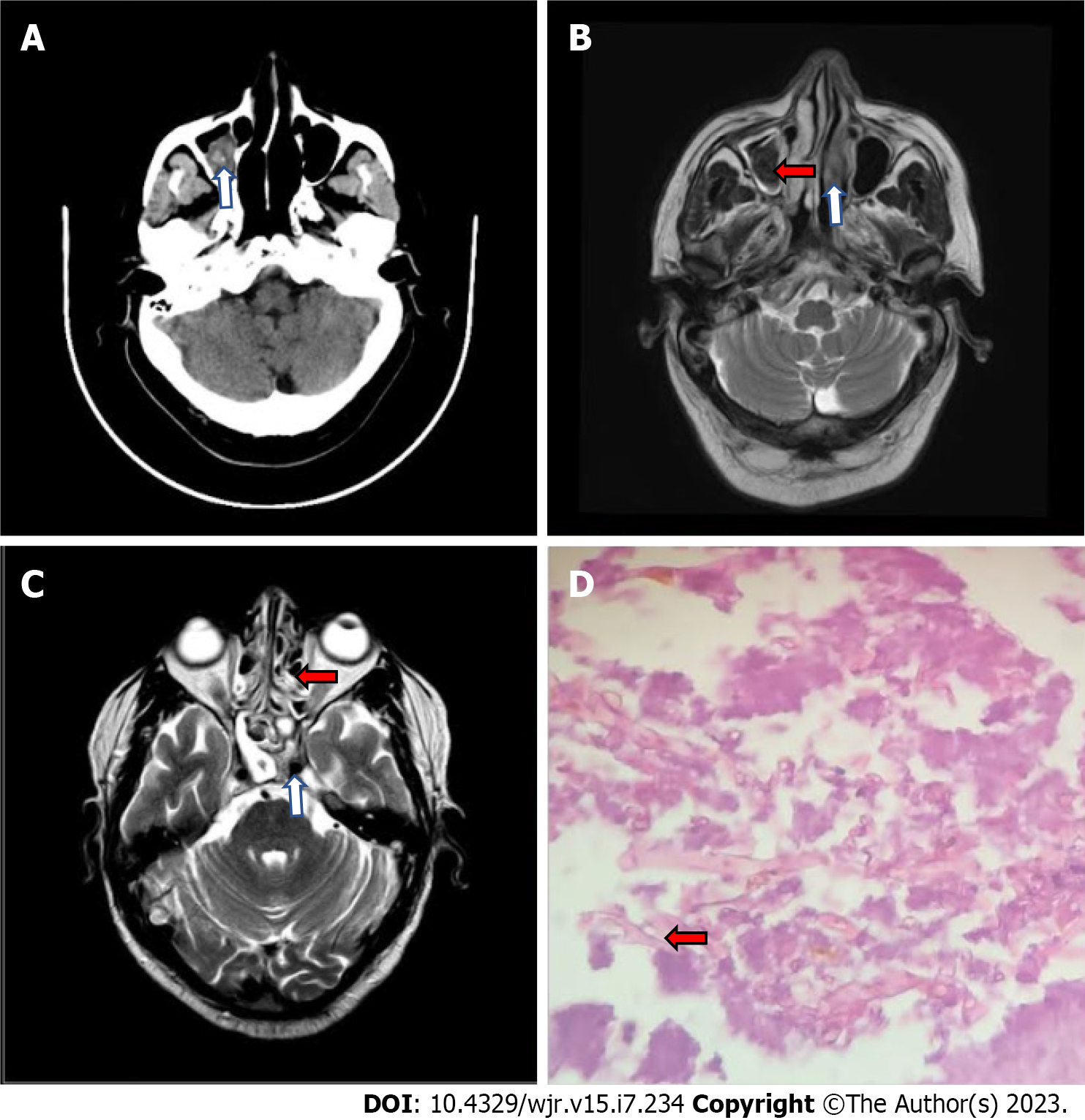
Figure 1 Images of patient with post COVID-19 mucormycosis.
A: Axial computed tomography image of brain, 10 d post admission demonstrating hyperdensity in right maxillary sinus (white arrow); B: Axial T2 weighted (T2W) and magnetic resonance imaging (MRI) image of brain demonstrating mucosal thickening with hypointensity within right maxillary sinus (red arrow) and hypointense left inferior turbinate (white arrow); C: Axial T2W and MRI image of brain demonstrating bilateral sphenoid and ethmoidal sinusitis (red arrow) with normal flow void within left internal carotid artery (white arrow); D: Histopathology slide demonstrating broad, aseptate ,branched hyphae of mucormycosis (red arrow).
- Citation: Narra R, Rayapati S. Invasive rhinocerebral mucormycosis: Imaging the temporal evolution of disease in post COVID-19 case with diabetes: A case report. World J Radiol 2023; 15(7): 234-240
- URL: https://www.wjgnet.com/1949-8470/full/v15/i7/234.htm
- DOI: https://dx.doi.org/10.4329/wjr.v15.i7.234


