Copyright
©The Author(s) 2023.
World J Radiol. Jun 28, 2023; 15(6): 157-169
Published online Jun 28, 2023. doi: 10.4329/wjr.v15.i6.157
Published online Jun 28, 2023. doi: 10.4329/wjr.v15.i6.157
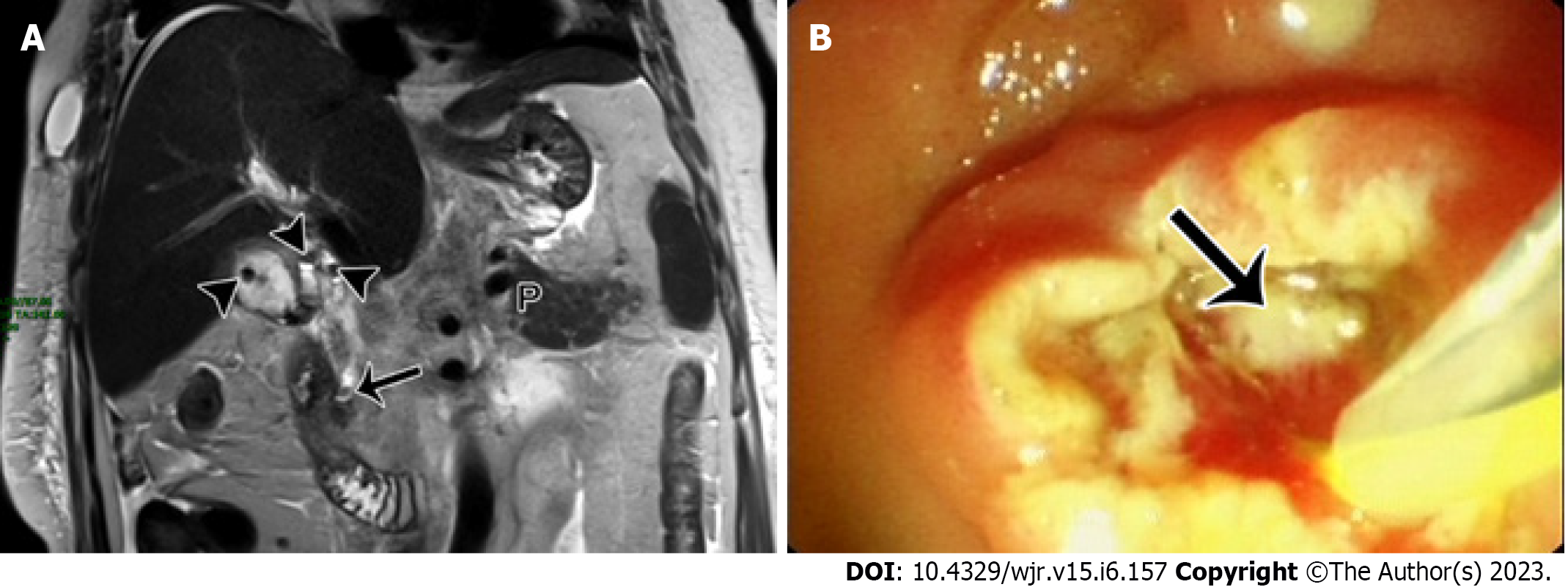
Figure 4 The magnetic resonance imaging report description needs to be focused on gallstone pancreatitis.
A 56-year-old woman with acute gallstone pancreatitis. A: Magnetic resonance imaging T2WI coronal imaging shows multiple hypointensity stones (arrowheads) in the gallbladder and gallbladder duct, and another hypointensity stone (arrow) in the lower level of the common bile duct. The patient was underwent an endoscopic retrograde cholangiopancreatography (ERCP) procedure; B: ERCP shows a stone in the lower part of the common bile duct with suppurative conditions (arrow). P: Pancreas.
- Citation: Song LJ, Xiao B. Acute pancreatitis: Structured report template of magnetic resonance imaging. World J Radiol 2023; 15(6): 157-169
- URL: https://www.wjgnet.com/1949-8470/full/v15/i6/157.htm
- DOI: https://dx.doi.org/10.4329/wjr.v15.i6.157


