Copyright
©The Author(s) 2023.
World J Radiol. Jun 28, 2023; 15(6): 157-169
Published online Jun 28, 2023. doi: 10.4329/wjr.v15.i6.157
Published online Jun 28, 2023. doi: 10.4329/wjr.v15.i6.157
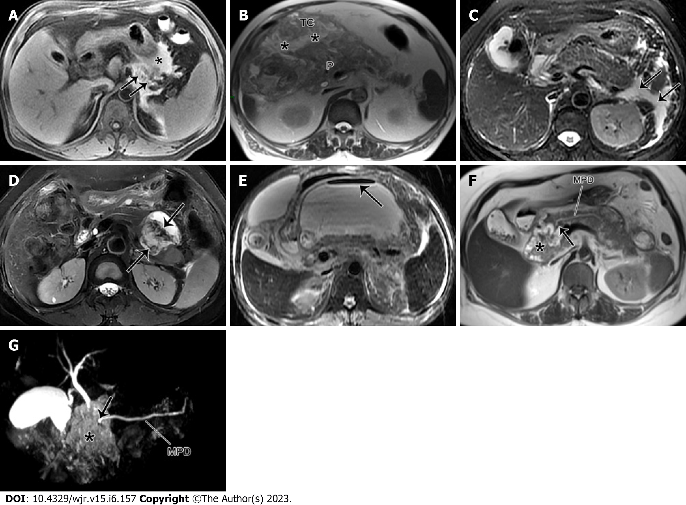
Figure 3 Pancreatic necrosis only.
A: A 47-year-old male with acute necrotizing pancreatitis complicated by hemorrhage. Magnetic resonance imaging (MRI) fat-suppressed T1WI depicts a large area of hyperintensity in the pancreatic body (arrow) and peripancreatic areas (*), indicating the presence of pancreatic and peripancreatic fat necrosis with hemorrhage; B: A 73-year-old man with acute necrotizing pancreatitis complicated with transverse mesenteric effusion. MRI T1-weighted imaging (T1WI) demonstrates peripancreatic inflammation spreading from the root of mesentery to the transverse colon along the involved transverse mesentery (*). P: pancreas; C: A 63-year-old woman with acute interstitial pancreatitis with acute peripancreatic fluid collection. MRI fat-suppressed T2WI reveals the uniformly hyperintense fluid collections (arrows) around the pancreas; D: A 53-year-old woman with walled-off necrosis secondary to acute necrotizing pancreatitis (pancreatic and peripancreatic necrosis type). MRI fat-suppressed T2WI shows an enveloped necrotic collection involving the body and tail of the pancreas, with solid necrotic debris (arrows) accounting for more than 40%; E: A 45-year-old man with acute necrotizing pancreatitis accompanied by walled-off necrosis and secondary infection. MRI fat-suppressed T2WI shows extensive walled-off necrosis in the omental sac, as well as a gas-fluid level sign (arrow). Thereafter, the open surgery and drainage for infectious collections was performed; F: A 55-year-old woman with pancreatic duct disruption syndrome secondary to acute necrotizing pancreatitis with walled-off necrosis. MRI T2WI shows an enveloped necrosis lesion (*) in the pancreatic head, and a cut-off sign (arrow) of the main pancreatic duct (MPD) traveling into this lesion (*). G: MRCP reveals that the MPD of the pancreatic body and tail directly enters into the lesion (*) in a right-angle, concomitant with the interrupted MPD.
- Citation: Song LJ, Xiao B. Acute pancreatitis: Structured report template of magnetic resonance imaging. World J Radiol 2023; 15(6): 157-169
- URL: https://www.wjgnet.com/1949-8470/full/v15/i6/157.htm
- DOI: https://dx.doi.org/10.4329/wjr.v15.i6.157


