Copyright
©The Author(s) 2023.
World J Radiol. Jun 28, 2023; 15(6): 157-169
Published online Jun 28, 2023. doi: 10.4329/wjr.v15.i6.157
Published online Jun 28, 2023. doi: 10.4329/wjr.v15.i6.157
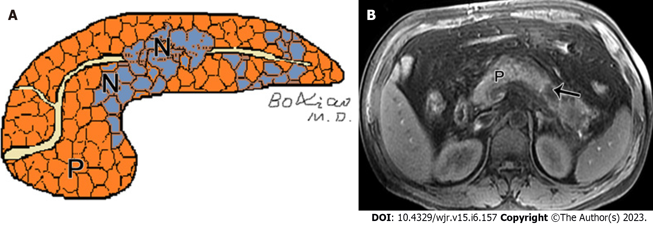
Figure 2 Peripancreatic necrosis only.
A: Schematic diagram of necrotizing pancreatitis (pancreatic necrosis alone): Scattered necrotic lesions (N) within the pancreatic parenchyma; B: A 40-year-old man with necrotizing pancreatitis (pancreatic necrosis alone). Magnetic resonance imaging fat-suppressed T1-weighted imaging shows hypointensity area (arrow) in the pancreatic body, as well as absence of peripancreatic fat involvement.
- Citation: Song LJ, Xiao B. Acute pancreatitis: Structured report template of magnetic resonance imaging. World J Radiol 2023; 15(6): 157-169
- URL: https://www.wjgnet.com/1949-8470/full/v15/i6/157.htm
- DOI: https://dx.doi.org/10.4329/wjr.v15.i6.157


