Copyright
©The Author(s) 2023.
World J Radiol. Jun 28, 2023; 15(6): 157-169
Published online Jun 28, 2023. doi: 10.4329/wjr.v15.i6.157
Published online Jun 28, 2023. doi: 10.4329/wjr.v15.i6.157
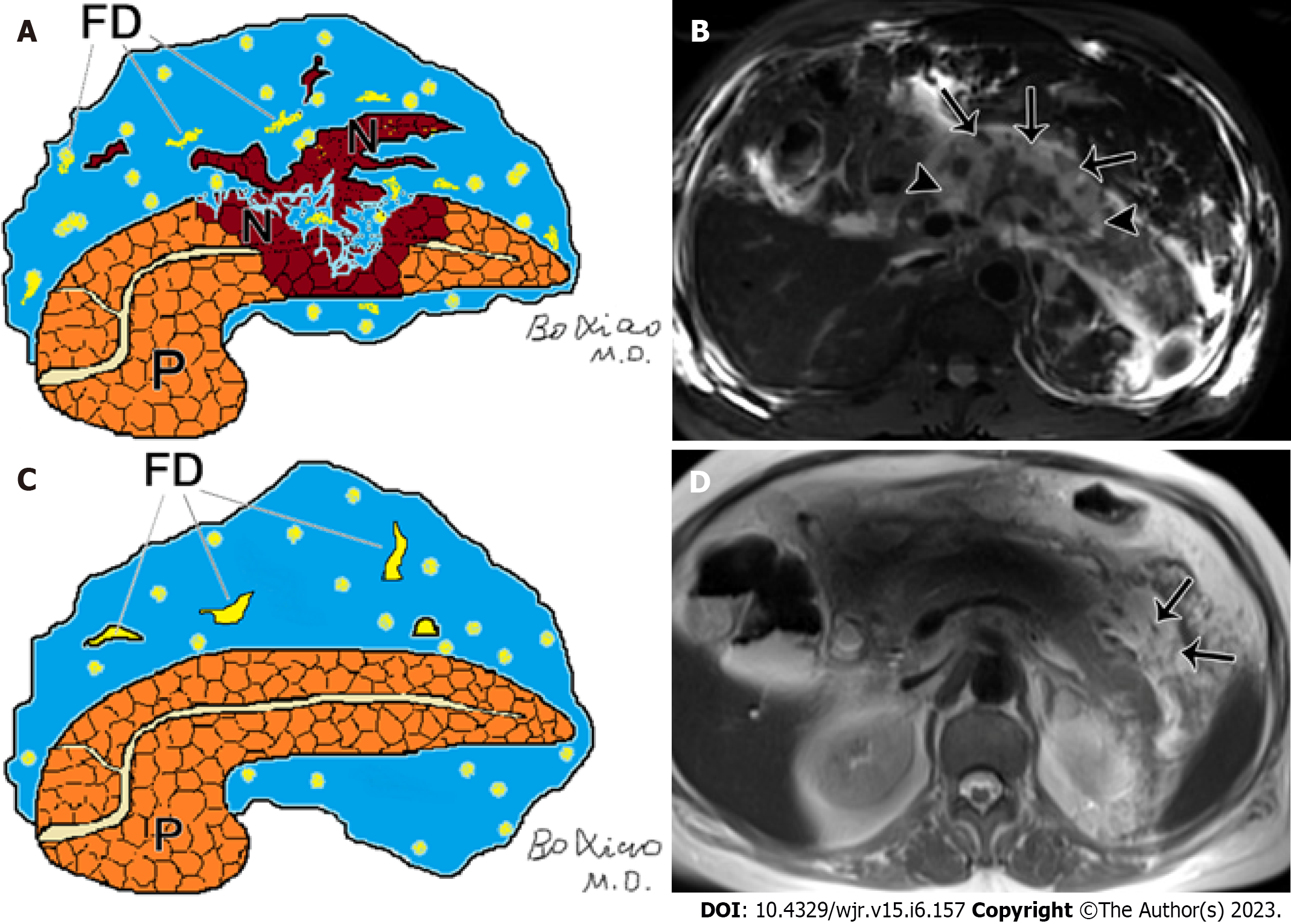
Figure 1 Pancreatic and peripancreatic necrosis.
A: Schematic diagram of pancreatic and peripancreatic necrosis (mixed type): pancreatic body necrosis (N) accompanied by peripancreatic fatty tissue debris (FD); B: A 61-year-old male with acute necrotizing pancreatitis (both pancreatic and peripancreatic necrosis). Fat-suppressed axial T1-weighted imaging shows a wide range of necrosis of the head and body of the pancreas (arrowheads), as well as peripancreatic collections containing large amounts of fat necrotic debris (arrows); C: Schematic diagram of necrotizing pancreatitis (peripancreatic necrosis alone): FD and absence of necrosis of pancreatic parenchyma; D: A 65-year-old woman with acute necrotizing pancreatitis (peripancreatic necrosis alone). Magnetic resonance imaging T2WI shows multiple patchy fatty fragments (hypointensity areas) (arrows) surrounding the pancreas.
- Citation: Song LJ, Xiao B. Acute pancreatitis: Structured report template of magnetic resonance imaging. World J Radiol 2023; 15(6): 157-169
- URL: https://www.wjgnet.com/1949-8470/full/v15/i6/157.htm
- DOI: https://dx.doi.org/10.4329/wjr.v15.i6.157


