Copyright
©The Author(s) 2023.
World J Radiol. Apr 28, 2023; 15(4): 98-117
Published online Apr 28, 2023. doi: 10.4329/wjr.v15.i4.98
Published online Apr 28, 2023. doi: 10.4329/wjr.v15.i4.98
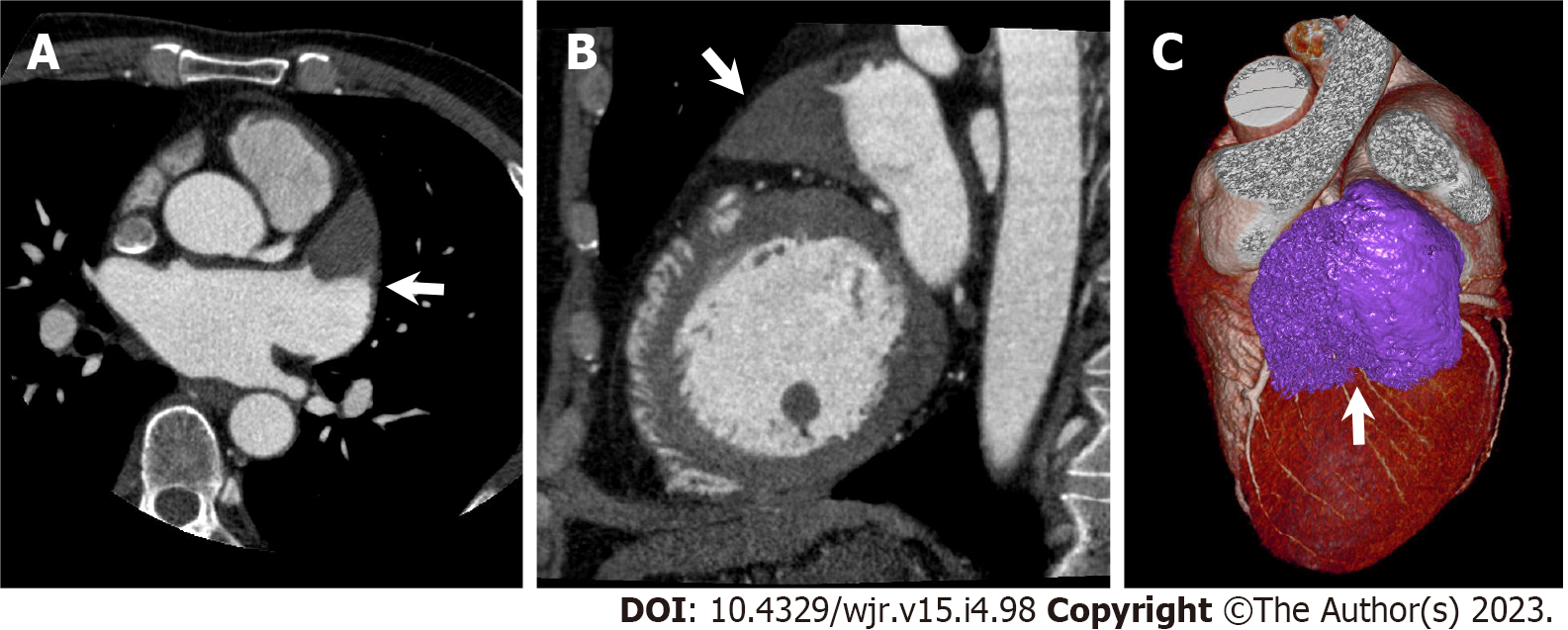
Figure 5 Left atrial appendage aneurysm.
A 56-year-old man with atrial fibrillation (Af) underwent cardiac computed tomography (CCT) to rule out obstructive coronary artery disease before catheter ablation for Af. A and B: Axial (A) and sagittal (B) reformatted CCT images show enlarged left atrial appendage (LAA, arrow) with triangular filling defect inside. LAA orifice diameter, body width, and length were 3.4 cm, 5.2 cm, and 6.8 cm, respectively; C: Three-dimensional volume-rendered image of CCT shows LAA aneurysm. CCT-derived maximum LAA volume was 56 mL.
- Citation: Yoshihara S. Evaluation of causal heart diseases in cardioembolic stroke by cardiac computed tomography. World J Radiol 2023; 15(4): 98-117
- URL: https://www.wjgnet.com/1949-8470/full/v15/i4/98.htm
- DOI: https://dx.doi.org/10.4329/wjr.v15.i4.98


