Copyright
©The Author(s) 2023.
World J Radiol. Mar 28, 2023; 15(3): 83-88
Published online Mar 28, 2023. doi: 10.4329/wjr.v15.i3.83
Published online Mar 28, 2023. doi: 10.4329/wjr.v15.i3.83
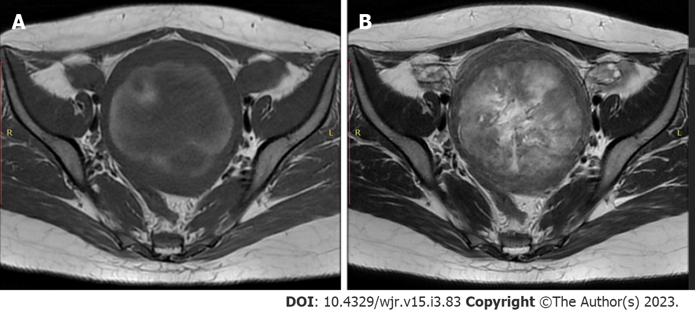
Figure 1 Magnetic resonance imaging findings.
An axial view T1 and T2-weighted showed a large intramural lesion in the myometrium with materials. A: High-signal intensity in T1-weighted view indicated the presence of a hemorrhagic component; B: High-signal intensity material in T2-weighted view indicated the presence of a liquid component.
- Citation: Martínez D, Sanchez GE, Gómez J, Sonda LJ, Suárez LD, López CS, Vega JJ, Cepeda DA. Magnetic resonance imaging findings of spontaneous pyomyoma in a premenopausal woman managed with myomectomy: A case report. World J Radiol 2023; 15(3): 83-88
- URL: https://www.wjgnet.com/1949-8470/full/v15/i3/83.htm
- DOI: https://dx.doi.org/10.4329/wjr.v15.i3.83


