Copyright
©The Author(s) 2023.
World J Radiol. Mar 28, 2023; 15(3): 69-82
Published online Mar 28, 2023. doi: 10.4329/wjr.v15.i3.69
Published online Mar 28, 2023. doi: 10.4329/wjr.v15.i3.69
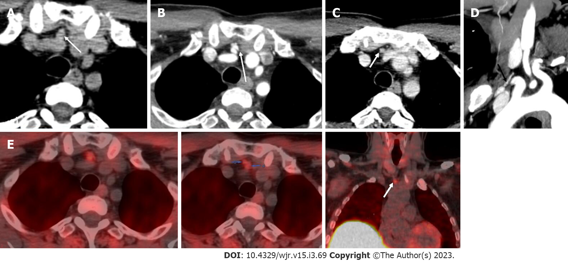
Figure 9 Ectopic parathyroid adenoma in the anterior mediastinum.
A: Computed tomography in a 45-year-old male patient showed a well-defined ovoid lesion in the prevascular space just posterior to the sternal notch and anterior to the inferior thyroid vessels appearing hypodense on the non-contrast phase; B: Showing intense enhancement on the arterial phase; C: washout in the venous phase; D: Coronal maximum intensity projection image demonstrates the inferior thyroid artery supplying the lesion (feeding vessel - black arrows); E: Fluoro-choline positron emission tomography shows a small tracer avid lesion in ectopic location which correlates with the computed tomography images.
- Citation: Gulati S, Chumber S, Puri G, Spalkit S, Damle NA, Das CJ. Multi-modality parathyroid imaging: A shifting paradigm. World J Radiol 2023; 15(3): 69-82
- URL: https://www.wjgnet.com/1949-8470/full/v15/i3/69.htm
- DOI: https://dx.doi.org/10.4329/wjr.v15.i3.69


