Copyright
©The Author(s) 2023.
World J Radiol. Mar 28, 2023; 15(3): 69-82
Published online Mar 28, 2023. doi: 10.4329/wjr.v15.i3.69
Published online Mar 28, 2023. doi: 10.4329/wjr.v15.i3.69
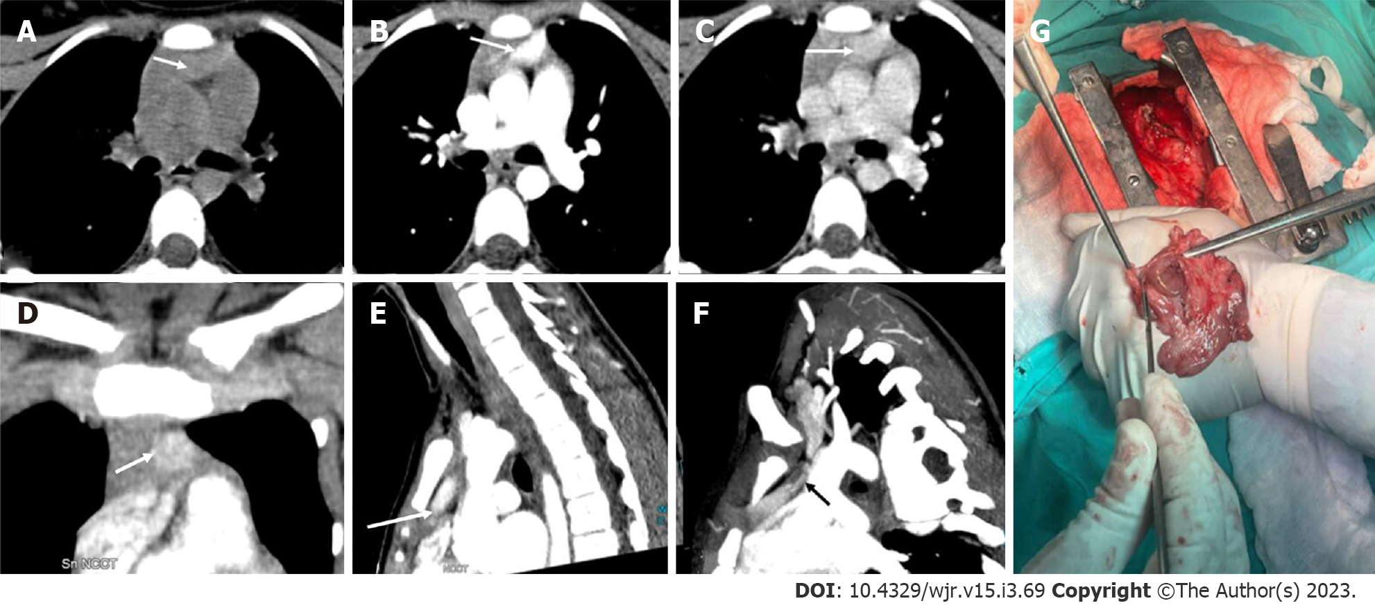
Figure 8 Ectopic parathyroid adenoma in the anterior mediastinum.
A: 4-dimensional computed tomography done in the 12-year-old female with hyperparathyroidism showed a well-defined lesion in the anterior mediastinum just behind the sternum which was hypodense on non-contrast computed tomography; B: intense arterial enhancement; C: washout on the venous phase; D: Multiplanar reformatted coronal; E: sagittal images in the arterial phase show the lesion better; F: Oblique sagittal maximum intensity projection image shows the feeding vessel (black arrow); G: Sternotomy followed by thymectomy was done and the thymus opened - the adenoma can be seen within the thymic parenchyma as pointed by the forceps.
- Citation: Gulati S, Chumber S, Puri G, Spalkit S, Damle NA, Das CJ. Multi-modality parathyroid imaging: A shifting paradigm. World J Radiol 2023; 15(3): 69-82
- URL: https://www.wjgnet.com/1949-8470/full/v15/i3/69.htm
- DOI: https://dx.doi.org/10.4329/wjr.v15.i3.69


